|
PARASITIC
SKIN INFESTATIONS
|
|
|
|
Leishmaniasis is a protozoal disease caused by Leishmania tropica parasite, which is transmitted by the Phlebotomus sand fly .The reservoir hosts are the dog in the Mediterranean area , man in the Middle East and the wild rodents in Asia and Africa . Leishmaniasis has three different morphological features ; cutaneous leishmaniasis , muco-cutaneous and the visceral (Kala-azar ).
Cutaneous leishmaniasis has many local synonym such as Tropical sore, Oriental sore, Aleppo sore or Baghdad sore. The disease is caused by Leishmania tropica protozoa, which is endemic in Asia minor, Southwest Asia, the Mediterranean and gulf regions. Modes of Infestations The Phlebotomus sand fly is the vector, transmitting the disease from the reservoirs to human being. Direct infection from infected sores to a traumatized skin may rarely cause the disease . Children are more susceptible, where solid immunity is acquired after the first infestation. This is why some natives sometimes inoculate their children with the protozoa on the shoulder or thighs to have the disease there in order to protect the face from scarring if they are infested in the future with leishmania. Clinical Features The disease has a very chronic course. The incubation period may take from weeks to two months from the beginning of the sand fly bite . Leishmaniasis usually affects children more than other age groups where the face , extremities and the neck are the most common sites involved.
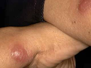
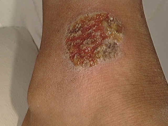
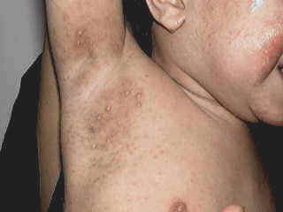
Cutaneous leishmaniasis has different clinical manifestations : Oriental sore The primary lesion is a papule mainly on the exposed areas such as the face and extremities . The papule enlarges after few weeks to form a round plaque, which later on ulcerates exuding a sticky secretion and forming a brownish , thick adherent crust .
Secondary bacterial infection of the ulcers is common causing more tissue destruction and disfiguring of the skin. One of the characteristic of tropical ulcer lesion is its long chronic course and the satellites, which develop nearby the primary lesion . These satellites may fuse together forming punched out rounded or oval ulcers that may heal after a few months causing disfiguring scars. Abortive type : Some lesions are dry where the papule changes into a nodule that may enlarge in size without ulceration .
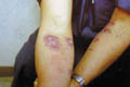
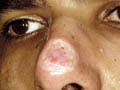
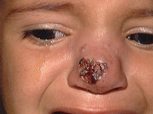
Different Clinical Manifestations Cutaneous leishmaniasis due to L. infantum : Infants infested with this parasite, may have visceral leishmaniasis, while adults usually get only the cutaneous lesions. Cutaneous leishmaniasis due to L. major: (This type is endemic in wet and rural zoonotic areas) The cutaneous lesions are red, furuncle-like nodule appears at the site of inoculation where after 2 weeks a central crust forms which may be followed by ulceration . The ulcer has a raised red margin , enlarges over the next 2-3 months where the lesion reaches a diameter of 3-6 cm. Multiple satellite nodules may develop near the primary lesion . Spontaneous healing even without treatment usually takes place within six months leaving a scar. Cutaneous leishmaniasis due to L. tropica The incubation period is more than 2 months . The lesion appears as a small, brownish nodule that enlarges gradually to a plaque 1-2 cm in diameter in about 6 months forming shallow ulceration with adherent crust. Secondary satellite lesions are minimal with this type . Cutaneous leishmaniasis due to L. Ethiopia Lesions are most commonly involve central of the face and theses are usually single. Satellite papules acuminate may form a large spreading nodule without crusts . Mucocutaneous lesions around the mouth and nose may occur . Diagnosis of Leishmaniasi
Differential Diagnosis Oriental sore may simulate different skin diseases such as tuberculosis cutis, tropical ulcer, tuberculus ulcer and others.
Mucocutaneous leishmaniasis is caused by Leishmania Brazilians parasite, which has the affinity to involve the skin and mucous membranes. The disease is endemic in Latin America, Peru and Brazil . Etiology The Phlebotomus fly transmitting the disease lives in the forests and causes the infection by biting their hosts . The parasite is present in two forms; the flagellate, which is found in the digestive tract of the vector and the non flagellate form which, is found in the tissues of human and animals infested by the parasite . Clinical Picture The incubation period is from 2-4 weeks. Skin manifestations The primary lesion is a nodule that may abort after few weeks or enlarge into a nodule which becomes vegetative and eventually forms a well-defined, irregular, infiltrating ulcer . Mucous membranes manifestations The characteristic of the mucocutaneous lesion is its tendency to metastasis to the mucous membrane of buccal and nasopharynex probably via lymphatics or blood stream. General manifestations The mucous membrane lesions may involve the adjacent cartilage while bones are spared . Disfiguring of the nose, soft palate, larynx, and pharynx . Ulceration of tongue, ocular and genital mucosa. Regional lymphadenopathy. Differential Diagnosis
Treatment of Leishmaniasis Most sores will heal spontaneously within one year . Treatment of cutaneous and muco cutaneous leishmaniasis is the same while the latter needs more intensive treatment due to the more severe and destructive complications. ** Pentavalent antimony: used for sores that may cause scarring and disfiguring on the face, lower leg or over a joint; mucosa or cartilage, or sores that might be due to parasites of the L. braziliensis. Unfortunately some cases of leishmaniasis, may be seen treated by topical steroid preparation. This changes the clinical picture, deteriorates the lesion , which becomes later more chronic and decreases its response to the specific medications. For adults, we give 6 cc of Pentostam I.M. daily for 10 days. This usually gives very good results, causing rapid healing of the ulcers. The dose is adjusted according to the age. ** El-Zawahry reported good results with dihydroemetine (Ciba) 2 tablets daily for adult age for one month ** Neostibosan (Bayer): is also an effective medication . The daily dose is 5mg./kg. body weight . A dose of 200-300 mg. can be given for older children and adults daily for 16 days is proved to be also effective . Other medications such as Chloroquine , Fouadin and antibiotics such as Tetracycline have been found to be effective. ** Pentamidine isethionate can be used for Leishmania tropica in a dose of 4 mg/kg body weight once weekly for as long as necessary . Patients with diffuse cutaneous leishmaniasis require treatment for a longer time. Leishmaniasis recidivans may respond to local infiltration, or systemic antimonies. ** Local infiltration with 1-2 ml sodium stibogluconate for solitary lesions. ** CO2 snow for small sores may be frozen and curetted under local anesthesia. Severe scarring lesions may require plastic surgery . ** We used Co2 laser to resurface and ablate cutaneous leishmaniasis lesions. In our medical center we recorded encouraging results especially in lesions complicated by scarring .
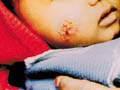
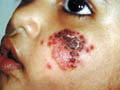
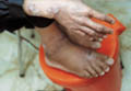
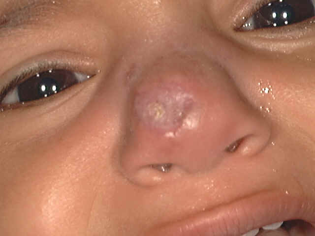
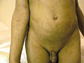
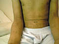
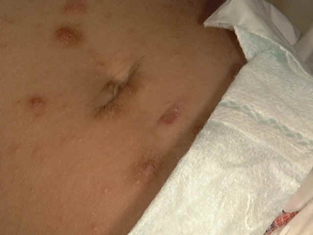
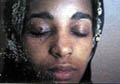
** Zithromx and Muperacin This child has cutaneous leishmaniasis since eight months. Different types of standard treatment even carbon dioxide and surgical debridement were tried in different centers.His father was advised for the last to be treated by Co2 Laser. I was not sure that is the best in such an area were scarring was clear due to different treatments. I prescribed for him muperacin cream (Bactroban cream and Zithromax suspension for one week) Dramatic relief of the lesion was noticed. The same line of treatment (Muperacin cream &Zithromax) was applied to other patients and the results were rappid healing of the lesion. Therefore , this safe , un expensive new line of treatment for cutaneous leishmaniasis can be tried. Fig.122 b. Cutaneous leishmaniasis (before treatment) Fig.122cOne week after treatment with Zithromax &Muperacin cream
Fig.117a. Oriental sore
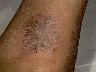
Fig.117b. Oriental sore *(One week after treatment with Muperacin cream (Bactroban) and Zithromycin uspension (Zithromax) . This is known also "Kala-azar" and "dum-dum fever." The disease is widely distributed in Asia, South Europe, around the Mediterranean, Africa, and in rural and poor communities . Clinical Picture Irregular fever of long duration .
POST KALA-AZAR DERMAL LEISHMANIASIS Clinical Features Cutaneous apple jelly nodules surround the healing scars mainly on the face may appear few years after healing of the primary lesions. Hypopigmented patches particularly on the face, neck, and extensor surface of the forearms and inner side of the thighs resembling lepromatous leprosy . Nodular and granulomatous lesion may appear on the skin and rarely Papillomatous on the eyelids, lips and ala nasi.
SOUTH AMERICAN TRYPANOSOMIASIS This is a systemic disease caused by Trypanosome cruzi. Modes of Infection Direct infection: Infection from infected animals . From animal reservoirs such as cats, dogs and wild rodents. Indirect infection : From man to man by the "kissing bugs" either, by the bug bite where trypanosomes are seeded into the skin, or infection from contamination with the bug feces . Human infection occurs chiefly through the skin and rarely through the mucous membrane . Clinical Features Infection is more severe in infants and young children . The primary stage: may be acute accompanied by constitutional symptoms such as fever, malaise and fatigue . Skin manifestations: severe reaction at the site of entrance of the parasite that may be in the form of erythema multiforme and edema . Late stage : This is due to hematogenous spread of the parasite to the viscera, the heart, brain and liver which may lead to serious complications and may be fatal .
SLEEPING
SICKNESS The transmitting vector is the tsetse fly which inocthe trypanosoma( (T. Rhodesian and T. gambiense) present in its salivary glands into the skin of the victim. Clinical Picture Acute stage : This stage is characterized by:
Skin manifestations : Chancre: nodule appears at the site of the bite, which is hot, red, tender and accompanied by lymphangitis and regional lymphadenopathy. Pruritus and painful edema involves the hands, feet and eyes. The joints become swollen. Late stage: Develops after a chronic course where there may be cerebral impairment that develops gradually leading to the clinical picture that is the "sleeping sickness". There is no skin manifestation in this stage . Diagnosis Detection of the parasites in the fluid aspirated from lymph nodes . Parasites are rare in blood .
Toxoplasmosis is a zonosis caused by the parasites, protozoon Toxoplasma gonadii. The disease is congenital, transmitted either from the infected mother to the fetus through the placenta or acquired from animal reservoirs such as cats , dogs and birds. Congenital Toxoplasmosis Infection of a pregnant woman may lead to abortion or delivery of a full term fetus with triad manifestations which are:
In subsequent pregnancies the fetus is not affected. Acquired Toxoplasmosis : The disease is contracted by contact with cats, rabbits, chicken, cattle and pigeons. Skin manifestations: Scarlitiniform eruption, urticarial, pinkish papules or subcutaneous nodules and rarely vesicles appear on the skin sparing the face, palms and soles. Systemic manifestations: multiple organ involvement causing encephalitis, hepatitis and other systemic manifestations. Diagnosis
Treatment Combination of Sulfonamides (adult dose 3g. daily) and pyrimethamine (Daraprim), given in a dose of 1mg/kg. daily for one month. It should be noted that Daraprim is a folic acid antagonist, so concomitant folic acid therapy is recommended. Rovamycin can give good results . Spiramycin (adult dose 2g. Daily ) is also effective . These medications reduce the incidence of fetal defects. When the proper medication is given to infected pregnant women. This drug can control the constitutional symptoms such as fever and improves the ocular lesions when combined with corticosteroids.
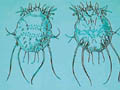
Zoonosis include the diseases that are caused by parasites. These parasites may be living on the skin surface (epizoonosis) or may pierce the epidermis (endozoonosis). The parasites may cause different skin manifestations : Scabies- Sarcopetes scabeii parasites cause itchy skin lesions due to their burrow into the skin. Bird mites infest birds such as chicken, canaries and other birds that may cause severe pruritic skin reaction. Some strains cause viral encephalitis when the mites attack human beings. Mouse mites cause skin reaction at the site of biting human beings and these also transmits rickettsia pox. Grain itch mite causes severe dermatitis due to the pediculoides mite. The disorder is called also "straw itch " . This occurs in families during the harvest time due to contact with the infested straw with the parasite.
Pulicosis is an acute epizoonosis due to fleabite . Pediculosis is a chronic epizoonosis , caused by lice . Cimicosis is a chronic skin reaction, caused by bed bugs Epizoonosis is a skin manifestations, due to insects such as wasps , bees and ants. Culicosis is due to mosquito bite causing pruritic macular and nodular lesion. The reaction may be severe in sensitized persons. In children the reaction may be severe causing papular urticaria, bullous reaction or reaction simulating lichen urticatus . Trombidiosis (hay or harvest itch ) is an acute epizoonosis that is caused by soil mite whose larvae may be attached to skin eliciting skin reaction in the form of erythematous papulovesicular lesions or exanthemas with excoriations and secondary bacterial infection such as furunculosis and impetigo. Creeping eruption is a skin disease that is caused by migrating nematodes during a stage of their development such as ancylostoma, hook worms, ascaris. The larvae migrate beneath the skin, forming burrows that cause pruritus and erythematous wheals .
SKIN
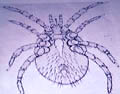
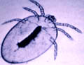
MANIFISTATIONS DUE TO MITES Human scabies is caused by the female parasite "Sarcopetes scabeii" which is capable of completing her life cycle in man. The female burrows into the skin after impregnation forming the characteristic lesion for scabies, which is known "the tunnel or burrow" where larvae are produced after eggs hatching. After copulation, larvae burrow into the skin and start again a new life cycle.
Modes of Infestation
Clinical Picture The disease has a chronic course. The incubation period may take weeks or even months without any apparent manifestations. The clinical picture varies according to age, sites involved and the type of host transmitting infestation . Scabies is characterized clinically by severe itching especially at night and when the skin becomes warm. Excoriation marks are due to severe scratching which may be accompanied by secondary bacterial infections in the form of impetigo or carbuncles. In the early stage, the burrows, where the Sarcopetes scabeii are impregnated into the skin can be seen easily as a grayish tortuous line where the mites are embedded at one site of the line. The sites infested by scabies have characteristic distribution. The commonest sites involved are the interdigital spaces of the fingers, the palms, the flexor surface of the wrests around the umbilicus, the region of the belt line, nipple, buttocks, genitalia and characteristically the glans penis in males. The face and neck are not involved except in infants .
Scabies transmitted from animals such as dogs and cats may have severe clinical picture such as macular , papular, pustular, impetigo or wheal like reaction . Itching is severe and may be distressing causing sleep disturbance.
Some neurotic patients who had scabies may continue to have a belief that the skin is still infested in spite of all the curative treatments he received. He considers any mild pruritus even insect bite is an attack of scabies. He moves from one clinic to the other and sometimes carrying with him in a container some skin debris to convince the doctor that he is still having the disease . In spite of that it is not easy to convince such patients. Alll the possible efforts should be considered in order to reassure him and to try all the possible clinical and psychic methods to get relief of such problem.
This is a rare type of scabies in which the clinical picture is more severe than the ordinary scabies. This type of scabies is found in poor communities, low sanitary conditions and in malnourished individuals . Clinical Features Crusted purulent lesions appear on the face and genitalia . Hyperkeratotic lesions appear on the palms and soles with subungual and nail dystrophy. Psoriasiform scaly lesions appear on the trunk and extremities .
The clinical features of scabies in infants differ in certain respects from the lesions that occur in older children and adults. Clinical Features Extensive distribution of burrows . Vesicular and Vesiculopustular lesions on the hands and feet are not uncommon, and bullous lesions have been described. Extensive eczematization is frequently present .
Fig.
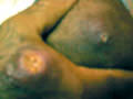
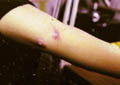
125b Scabies in babies
Fig. b. Scabies in babies (Papulovesicular and bullous lesions)
Fig.126 b. Scabies in babies (Papulovesicular and bullous lesions) There may be multiple crusted nodules on the trunk and limbs. Diagnosis of Scabies
The mite can be detected either by superficial pricking and raising the end of the burrow with 25 gauge needle, where the mite may be seen sticking to the edge of the needle. This can be detected also by very superficial shaving of the burrow and examining the materials shaved under the low power microscope. Treatment General measures The whole body, except the head and neck should be treated. All members of the family and close contacts should be treated, whether they are symptomatic or not. Treatment of scabies is easy if the patient follows the instructions and to use the medications in the proper way besides measures to prevent re-infestation . When scabies is suspected in any patient it is wise to begin treatment of this case as scabies until the diagnosis is confirmed. A pruritic skin disease whether scabies or not may benefit from the medications used for scabies where these can help at least to stop itching which is sometimes a big problem for patients and may interfere with sleeping. In children the mother is instructed to apply medications (Eurax or Kwell Lotion) to any part of the body involved especially the intertriginous and between the interdigital spaces of the fingers. The lotion or cream is left on the skin and washed next day using newly washed clothes preferred ironed on both sides . Boiling and ironing the clothes from both sides is usually enough to eradicate the mites. Some clothes may be damaged by boiling, dry cleaning and the usual method for laundery for the bed linen may be also sufficient to kill the mite. Disinfestations of clothing and bedding, other than by ordinary laundering, is not always necessary. Specific treatment Benzyl benzoate is available in concentrations of 25 per cent. This should be diluted with 2 or 3 parts water for use to infants and young children. Prolonged or repeated applications of Benzyl benzoate or Lindane should be avoided. Treatment of scabies is usually effective by rubbing the skin from neck to toe by Kwell lotion or Eurax after taking a hot bath and rubbing the skin with a sponge . Crotamiton lotion and cream may be used to treat burrows on the head and neck. The adverse effects of these medications may be contact dermatitis and toxic epidermal necrolysis attributed to Monosulfiram have been reported. Antihistamines are necessary to stop itching. Antibiotics orally may be needed to control secondary bacterial infections. Erythrocin, Cephalosporin or Fluxacillin orally are effective in controlling carbuncles or pustular lesions that complicate some cases of scabies.
SKIN MANIFESTATIONS OF OTHER MITES
The eruption provoked by these mites is commonly composed of minute, intensely pruritic papules or papulovesicles on the exposed parts of the body, principally on the head, neck, and forearms but occasionally more widespread. The appearance of the eruption on the face may suggest an acute contact dermatitis. Dermatophagoides pteronyssinus (the house-dust mite) is widely distributed in the human environment especially in house dust and beds. It occurs worldwide and has been reported from all inhabited continents. The largest numbers of mites are found in houses that are damp and inadequately heated. Numbers vary seasonally, increasing in early summer to reach a maximum by early autumn. The main food of D. pteronyssinus is human skin scales. Xerophylic moulds, especially Aspergillus Penicilloides, are essential for the growth and survival of D. pteronyssinus. The moulds digest lipid in the scales that is toxic to the mites.
The role of the house-dust mite in the pathogenesis of atopic eczema remains controversial. The houses of patients with moderate to severe atopic eczema had more house-dust mites than normal individuals. However, convincing direct evidence that house-dust mite exposure exacerbates atopic eczema is lacking. Measures to reduce house-dust mite numbers include regular vacuum cleaning of bedroom carpets, mattresses, and the use of plastic mattress covers. The antifungal drug "Natamycin " can be considered in the treatment. These types of mites attack infants and children in their beds, which may elicit or exacerbate dermatitis and allergic bronchial asthma in some patients. Skin tests can detect these mites. Skin desensitization may be of help for some patients.
PYEMOTES MITES Skin lesion is due to contact with infested straw . The dermatitis has been known as "barley itch," "grain itch," "straw itch," "cotton-seed dermatitis." The lesions are urticarial papules surmounted by vesicles and occasionally bullous. They are often very numerous, and their distribution depends upon the mode of exposure.
Different members of these mites are distributed worldwide. The most important of these mites are the red bug, which live in grasses, shrubs and even in houses. They are present in crowded places as refugee , military camps and prisons. The parasites attack human skin sucking their blood and fall down when the body is engorged . Infants and young children may have more severe skin reactions due to their delicate skin mainly on the legs , belt site , face and other areas of the body . Clinical Features Skin manifestations Itchy papules appear at the site of the parasite bite . These may enlarge to form a nodule . Systemic manifestations Some species of red bugs may be a vector for systemic diseases such as Tsutsu gamushi fever.
HARVEST MITES Dermatitis is due to the parasitic larvae, or free-living nymphs. The larvae may cause troublesome dermatitis . Some of these mites are important vectors of Rickettsial disease. The eggs are laid in soil. The six-legged larvae that emerge climb onto low vegetation to wait for suitable vertebrate hosts. On the host, the larvae move to areas where the skin is thin, such as the ears, axillae, groins and genitalia. They pierce the skin with their claws and inject saliva, which has cytolytic properties into the epidermis and feed on fluids and cell debris. Once engorged, they fall to the ground. The larval mites are most numerous from May to October, with a peak in September. The most favored natural host is the rabbit. Eutrombicula are the most common chiggers attacking man . Clinical Picture Infestation to children occurs while playing on grassy areas or whilst walking bare footed through grass or low vegetation. The response to the bites of harvest mites appears to be determined by the irritant effect of the mites‘ saliva and an acquired hypersensitivity to salivary antigens. Erythematous macules appear at the sites of the bites. Later on these gradually develop into extremely itchy papules or papulovesicles. In heavy infestations the lesion may cover extensive skin sites . The distribution and the type of lesion is determined by the preference of mites for thin skin as that of the crural areas of young children besides the type of clothing of the host. Lesions commonly occur around the feet and ankles, the groins and genitalia, the axillae, the wrists , antecubital fossa, and areas constricted by clothing, such as the waistline.
Species of Cheyletiella mites are non-burrowing. Obligatory parasites of certain mammals, predominantly in dogs, cats and rabbits. The entire life cycle is completed on the host. Skin lesions manifest when there is contact with infested animals. Clinical Features The typical clinical picture is large numbers of intensely itchy papules surmounted by a vesicle. Infection occurs on the areas coming in contact with the infested animals during fondling of these animals. These lesions become necrotic and the eruption may become extensive covering the chest and abdomen manifesting with severe itching. Secondary bacterial infection may complicate the skin lesions due to severe scratching .
DERMANYSSID MITES Dermanyssid mites are hematophagous parasites of birds and mammals. Dermanyssus gallinae (the poultry mite), is a common parasite of domestic and wild birds. Infestation is by playing or dealing with domestic birds such as pigeons, chicken or their cages. Mites enter houses from bird‘s nests via windows or air conditions causing dermatitis . Liponyssoides sanguineus, the house mouse mite, is an Ectoparasite of small rodents. It is of medical importance because it is the vector of Rickettsia akari that transmits Rickettsia disease.
Members of the Macronyssidae are hematophagous Ectoparasite of birds, mammals and reptiles. Clinical Picture The clinical manifestations vary according to the route , severity of infestation and the degree of the host‘s response. Skin manifestations are pruritic papular or urticarial lesions . A profuse eruption of small, intensely itchy wheals or papules appear which are sometimes grouped, and often asymmetrical. The lesions may have a central punctum, and vesicles occasionally occur in the center of the papules, especially in children. Secondary infection may occur due to severe itching. Persons attacked by mites in bedding have more extensive bites. Occasionally lesions are grouped adjacent to areas of tight clothing around the waistline. In heavy infestations the causative mites may be detected at the site of the parasite bite .
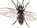
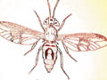
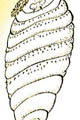
SKIN MANIFISTATIONS DUE TO FLIES Flies are an important vector of certain infectious diseases . Skin manifestations are either due to the adult fly bite or due to the larvae within the skin causing skin myiasis . Clinical Picture
The severity of manifestations vary depending on to the type of the insect family : Family Tabanidae: This is a fly animals such as horses and deers that causes a severe painful bite. Family Helediae: Known also as "no see ums" because of their extraordinarily small size. These have the characteristic of savage biting and cause itchy and irritable lesions that may last for a few days.
Family Simuliidae (black flies) - are endemic in temperate and suburban areas. These are dangerous and curious flies . Clinical Manifestations Black flies - cause painful bite, preferring the eyes, nostrils and ears. They cause severe local and systemic manifestations. Systemic manifestations - fever and gastrointestinal problems . Local reaction: an erythematous and edematous lesions appear, swelling of the face and may cause ulceration and distortion. Family Psychodidae: Species of the genus Phlebotomus are the vectors of cutaneous leishmaniasis and kala-azar, Carrion‘s disease, Verruga peruana and others. Phlebotomus attack their victims at night and prefer the ankles, wrist, knees and elbows. Family Chloropidae. This group is usually endemic in rural and urban areas with decreased sanitary care. They feed on human blood and eye secretions causing epidemic conjunctivitis , sores and open wounds.
Clinical Manifestations
Treatment of Myiasis Surgical extraction of the larvae with a sharp needle and douching of the wound with 15 per cent chloroform in vegetable oil . Anaesthetizing the larva using cotton pad moistened with Chloroform can treat nasal myiasis and then blocking the nostril for 2-3 minutes. Remove the larvae with forceps. Preventive measures by eradication of screw worm fly.
|


| Contents | << Previous Chapter | Next Chapter >> | Search |