CHAPTER 32

GENODERMATOSES

Contents << Previous Chapter Next Chapter >> Search

  
Recessive epidermolysis bullosa

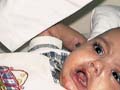
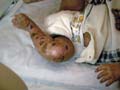
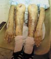
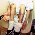
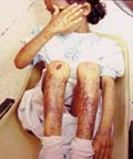
The recessive dystrophic epidermolysis bullosa has the same cutaneous features of the dominant type.

Skin manifestations

Large, flaccid often-hemorrhagic bullae appear after birth or early in infancy.

Mucous membrane manifestations

Severe scarring of the mucous membranes of the mouth, pharynx, esophagus and the condition may be fatal.

Nail dystrophy is a common complication of the recessive type of epidermolysis bullosa.

Secondary bacterial infection is common and causes more complications.


Fig. 256. Epidermolysis, Recessive 
Dystrophic Type 


Fig. 257. Epidermolysis Bullosa 
(Bullae & skin scarring)

Differential Diagnosis

  1. Bullous diseases
    In infancy, differentiation of epidermolysis bullosa from other bullous eruptions is rarely a problem. Benign chronic bullous disease of childhood may mimic inverse types of EB since the blistering occurs around the genitalia, perineum and buttocks. However, histology of these conditions is diagnostic and the presence of linear IgA on direct immunofluorescence may help to confirm the diagnosis.

  2. Dominant bullous ichthyosiform erythroderma.
    This type presents with flaccid bullae, but erythroderma and hyperkeratosis, especially over skin folds, may help in the diagnosis.

  3. X-linked incontinentia pigmenti is characterized by streaky blistering which results in swirly patterns of pigmentation especially over the trunk.

  4. Scalded skin syndrome

    Sudden onset of generalized erythema, fever and necrotic peeling of the epidermis is characteristic of staphylococcal scalded skin syndrome.

    Tinea, pompholyx and infective dermatitis diseases may be confused in children with the localized simple forms of epidermolysis bullosa.

Treatment

Treatment of recessive epidermolysis bullosa is not always curative.

High doses of steroids were tried and antibiotic to combat secondary bacterial infection.


GARDNER‘S SYNDROME

This syndrome is an autosomal dominant disease characterized by:

Multiple epidermoid cysts .

Congenital hypertrophy of the retinal pigmented epithelium

Clinical Features

Skin manifestations:

‘Sebaceous‘ or epidermoid cysts may appear in early childhood, which may be numerous. These are usually irregularly distributed on the face, scalp, extremities, and are less frequent on the trunk.

Cutaneous and skeletal changes may be present without polyposis and polyposis may be present when one or more of the other features of the syndrome is lacking. 

Lipomas in the subcutaneous tissues, and in other organs, have frequently been noted. Fibromas or desmoid tumors and fibrosarcomas are less frequently present.

Gastro-intestinal manifestations:

Polyposis of the colon or rectum usually arises during the second decade, but may occur in early childhood. Leiomyomas of the stomach or ileum and malignant changes develop later. Fibromatous growths of the mesentery may be discovered at operation and severe peritoneal scarring may follow surgery.

Bone changes

Osteomas are usually multiple that develop mainly in the maxilla and sphenoid bones and to a lesser extent in the long bones.

Treatment

Colonoscopy examinations, where the possibility of prophylactic colectomy should be considered in the second decade.


ECTODERMAL DYSPLASIA 

The ectodermal dysplasia is a genetic heterogeneous group of disorders with a primary defect in hair, teeth, nails or sweat gland function.

A. Hypohidrotic Ectodermal Dysplasia

(Christ-siemens-touraine syndrome)

(Anhidrotic ectodermal dysplasia)

The main features of the syndrome are:

Hypohidrosis

Hypotrichosis

Hypodontia.

Clinical Manifestations

Reduced sweating. Absent or reduced sweating causes heat intolerance and affected individuals may present with unexplained fever in infancy or childhood. Extreme discomfort can follow exertion or eating hot foods. 

Teeth manifestations : Total or partial anodontia are the essential features of the syndrome. The temporary and permanent teeth may be entirely absent, or there may be a few teeth present. The incisors and/or canines are characteristically conical and pointed. The jaws develop normally even in complete anodontia but the gums may be atrophic.

The conical, pointed teeth are the key feature of the syndrome and may be the only obvious abnormality. In other cases they may be associated with congenital alopecia, and defective sweating, if present, may be detectable only on appropriate testing.

Musculo-skletal abnormalities

Prominent frontal ridges and chin.

Saddle-nose.

Sunken cheeks.

Thick, everted lips

Large ears.

Skin manifestations

The skin is smooth, soft, and dry, finely wrinkled (especially around the eyes) and appears prematurely aged.

Mucous membranes manifestations

The mouth may be dry from hypoplasia of the salivary glands and the lacrimal glands may be deficient.

Atrophic rhinitis is common and some patients have no sense of smell or taste.

Internal organs manifestations

Poor development of mucous glands of the respiratory and gastrointestinal tract may increase susceptibility to respiratory infection, dysphagia, stomatitis and diarrhea.

Aplasia or hypoplasia of the breasts is occasionally noted. 

Hair Manifestations

Alopecia is often the first feature to attract attention, but it is seldom total. The scalp hair is sparse, dry, and fine and usually remains short. The structure of the shaft may be abnormal. The eyebrows are sparse or absent, but the lashes are usually normal. The beard, pubic and axillary hair is often sparse and other terminal hair on trunk and limbs may be absent.

Nail changes

The nails are abnormal in about half the cases and may be brittled, thin or ridged, but are seldom grossly deformed.

Eye manifestations

Ocular abnormalities such as corneal and lenticular opacities may occur.

Atopic eczema and asthma are common manifestations.

General physical development: may be somewhat stunted, but sexual development is usually normal. Primary hypogonadism is occasionally associated with the syndrome.

Mental development: Mental retardation appears in some cases. The expectation of life is normal or only slightly reduced.

Treatment

Restriction of physical exertion.

Choice of suitable occupation.

Avoidance of warm climates.

Special schooling

Psychological support may be needed.

Regular dental supervision is essential at an early age.

B) Eec Syndrome

This is an autosomal dominant syndrome characterized by ectrodactyly (lobster-claw deformity of the hand) with cleft lip and palate.

Clinical Manifestations

The main features of the EEC syndrome are:

Ectrodactyly

Sparse wiry and hypopigmented hair

Peg-shaped teeth with defective enamel

Cleft lip and palate.

Lacrimal duct stenosis.

Corneal scarring and blindness are serious complications of the syndrome.

The normal mucosal covering of the laryngeal folds is absent, and the voice tends to have a breathy quality, as in anhidrotic ectodermal dysplasia. Not all the defects are present in every patient.

Sweating may be normal in some patients. 

C) Rosselli-Gulienetti Syndrome

The inheritance of this syndrome is probably determined by an autosomal recessive gene.

Clinical Features

Hypohidrosis accompanies slight frontal bossing and some depression of the nasal bridge.

The scalp hair is often fine, dry, sparse and light in color, the nails are dystrophic and teeth are few and small. Other features are cleft lip and palate, syndactyly and defects of the external genitalia.

There may be popliteal web formation.

D) Alopecia-onychodysplasia- hypohidrosis Syndrome

This is a distinct syndrome of an autosomal recessive inheritance.

Clinical Manifestations

Hair

A child may be born bald and remain so, apart from the few thin yellow hairs on the scalp.

Nails : Thick toenails and the fingernails are normal.

Hyperkeratosis of the palms and soles knees and elbows and, to some extent, on the skin generally except on the head and neck.

Deafness may affect some cases and defects of other organs.

Teeth are small.

Hypohidrosis.


SYNDROMES ASSOCIATED WITH DNA INSTABILITY

Certain genetic syndromes have been grouped together because they share some features such as:

Chromosomal instability.

Hypersensitivity to irradiation or mutagenic chemicals.

The cellular DNA (i.e. the genome) can be damaged by irradiation, or by mutagenic or carcinogenic chemicals in the environment. Normal individuals possess the ability to repair this damage. In some diseases, the ability to repair damage to the genome seems to be impaired.

Increased risk of malignancy.

These diseases include: Xeroderma pigmontosum, Bloom‘s syndrome, Cockayne‘s syndrome, dyskeratosis congenita, progeria, Fanconi‘s anemia and ataxia telangiectasia.


XERODERMA PIGMENTOSUM

Xeroderma pigmentosum (XP) is a rare autosomal recessive disease, first described by Kaposi in 1870, characterized mainly by photosensitivity to wavelengths 280 to 310 nm, pigmentary changes, premature skin aging, neoplasia and abnormal defect in DNA repair.

Clinical Features

Freckles: varying in color and size, appear on the sun exposed areas first on the face and hands and later on other exposed parts, the neck and the lower legs, the lips and the conjunctiva where in severe cases the trunk is affected. The entire face mainly around nose and eyes shows pigmented spots of various tints of brown, mingled with white atrophic patch and telengectasia.


Fig.258. Xeroderma pigmentosum


Fig. 259. Xeroderma pigmentosum
(Freckles & photosensitivity)


Fig. 260. Xeroderma pigmentosum
(Freckles & photosensitivity)

Vesiculobullous lesions and superficial ulcers, healing with difficulty, leave disfiguring scars, and contractures that may produce ectropion and obliterate the outline of the eyelids. Kerato-acanthomas and actinic keratoses may undergo malignant change.


Fig.261. Xeroderma pigmentosum
(Freckles & photosensitivity)

Actinic keratoses, melanomas and squamous cell carcinoma are later manifestations.

Telangiectases and angiomas on unexposed skin and on the lingual and buccal mucous membrane have been reported. Small, round or irregular white atrophic spots are common late manifestations.

Ocular Lesions

The eyes are affected in most cases. Photophobia and conjunctivitis are common early symptoms. Ectropion and destruction of the lower lids expose the bulbar conjunctiva and synblepharon and ulceration may occur. Pigmented macules on the conjunctiva are common. Vascular pterygium, corneal opacities and epitheliomas of the lids, conjunctiva or cornea may develop.

Malignant Changes

The first malignant tumors may develop as early as the third or fourth year. Basal-cell carcinoma is common and large numbers, sometimes pigmented, may appear over the course of years. Squamous-cell carcinoma is also common.

Melanomas arise and may be multiple; they may lead to early death from widespread metastases.

The disease is often fatal before the age of 10, and two-thirds die before 20.

Neurological Manifestations

Neurological abnormalities occur in approximately 20% of XP patients with one or more of the following: mental retardation, areflexia or hyporeflexia, spasticity, ataxia, deafness, dysphasia.

De Sanctis-Cacchione syndrome

This term has been applied to the association of XP with microcephaly, severe mental deficiency, dwarfism, hypogonadism, deafness, choreoathetosis and ataxia. Post-mortem findings show cerebral and olivopontocerebellar atrophy from neuron loss, without primary damage to white matter, or gliosis.

Treatment of Xeroderma

General measures

Avoid exposure to sunlight.

Patients must not go outdoors during daylight hours, except in the early morning or evening, and even then they should wear two layers of clothing and a broad-brimmed hat. All uncovered skin surfaces must be protected by a total sun-block cream, and sunglasses with side shields should be worn. The UVR is harmful up to at least 320 nm, and some fluorescent lights can emit radiation below this wavelength.

The relatives of known cases should be carefully examined and tested so those mildly affected individuals may be detected at the earliest possible stage.

Treatment of lesions

Surgical

Early and adequate excision of all tumors is essential and it is to be preferred to radiotherapy on account of the atrophic and degenerate state of the skin.

Plastic surgery and grafting of large areas of facial skin may sometimes be required.

Topical medications

Topical 5-fluorouracil or dermabrasion may be useful for early or premalignant lesions.

The eyes may need to be treated with artificial tears, soft contact lenses or even corneal transplant.

Systemic medications: oral retinoids may be of help for some cases.

Pigmented Xerodermoid

The onset of pigmented xerodermoid may be delayed until the third or fourth decade. In pigmented xerodermoid, repair replication is normal, but there is almost total depression of DNA synthesis after exposure to UVR where patients must be protected from sunlight by every possible means.

Repair in this type is not impaired but there is almost total depression of DNA synthesis after exposure to UVR. This type appears in older age groups and the manifestations are delayed may up to the third or fourth decade.


POIKILODERMA CONGENITA
(Rothmund-Thomson Syndrome)

Rothmund-Thomson syndrome is a rare autosomal recessive, hereditary disorder, occurring predominantly in females, characterized by cataract and skin degeneration.

Clinical Manifestations

The skin appears normal at birth. The earliest lesions usually develop between the third and sixth month but sometimes as late as the second year.

Skin manifestations

Skin lesions begin as tense erythematous and edematous plaques on the cheeks, hand, feet and buttocks followed by fine or punctate reticulated atrophy, telangiectasia, pigmentation, depigmentation and ultimately, the lesions closely resemble chronic radiodermatitis.

Hair manifestations

Scalp hair is often sparse, fine, and may be absent. Eyebrows and lashes, pubic and axillary hair are often sparse or absent.

Teeth changes

Teeth are often normal, but microdontia and early caries have been reported.

Nails are normal, or small and dystrophic.

Photosensitivity on sun exposed areas, which may be severe eliciting bullous reaction.

Ocular manifestations

Bilateral cataracts, usually between the fourth and seventh year.

Neurological manifestations

Some cases show mental retardation, while physical development is frequently retarded and the affected patients are dwarf.

Endocrine changes: hypogonadism and hyperparathyroidism.

Mental development is usually normal, but mental retardation may develop in some cases.

Expectation of life appears to be normal.

Diagnosis

Age of onset: is one of the essential features in differential diagnosis.

Distribution of skin lesions mainly on the sun exposed areas.

Skin lesions are characterized by atrophy, telangiectasia and mottled pigmentation, most intense on light-exposed skin.

Photosensivity is a feature of the syndrome.

Differential Diagnosis

  1. Werner‘s syndrome
    The skin changes are essentially sclerodermatous, and both skin and ocular lesions develop later than in the Rothmund-Thomson syndrome.

  2. Dyskeratosis congenita
    Reticulate pigmentation develops between the ages of 5 and 13, and is most marked on the neck, trunk and thighs. Atrophy and telangiectasia may appear later. The nail changes are constant and severe.

  3. Progeria
    The child is often small, but otherwise normal during the first year, thereafter development is retarded. The scalp hair, brows and lashes are lost and the skin assumes an increasingly senile appearance.

  4. Cockayne‘s syndrome
    Light sensitivity is a feature after the first year, but there is no poikiloderma.

  5. Hypohidrotic ectodermal dysplasia.
    Conical teeth, hypotrichosis and partial or complete anhidrosis are the main features. The skin lesions show neither telengectasia nor pigmentation

  6. Xeroderma pigmentosum
    In mild forms only freckle-like macules are present.

  7. Rothmund-Thomson syndrome: Telangiectasia is the conspicuous feature.

  8. Focal dermal hypoplasia: Telangiectasias is often irregular, linear and present at birth.

  9. Bloom‘s syndrome: Erythema, and not poikiloderma, is the essential change.

Treatment of Poikeloderma 

Protection against sunlight is important and early diagnosis of any malignancy.

Retinoids may help some cases.


MARINESCO-SJO"GREN SYNDROME

The inheritance of this rare syndrome is determined by an autosomal recessive gene.

Clinical Features

Cerebellar ataxia is apparent as soon as the child begins to walk.

Dysarthria.

Mental and physical development is retarded.

Congenital cataracts associated with rotary and horizontal nystagmus.

Skeletal defects are commonly present.

The teeth are malformed and the lateral incisors may be absent.

The nails are thin and fragile.

The hair is sparse, fine, short, fair and brittle.


SECKEL‘S SYNDROME
(Bird-Headed Dwarfism)

The syndrome is a rare inherited by an autosomal recessive gene.

Clinical Features

Main features:

Prominent beak-like nose

Growth retardation, microcephaly and mental deficiency.

Other manifestations:

Hypoplastic face with large eyes.

Skeletal defects are frequent.

The hair may be sparse and prematurely gray.

Diagnosis

The syndrome must be differentiated from others in which there is intrauterine dwarfism.


CARTILAGE-HAIR HYPOPLASIA SYNDROME
(Metaphyseal Chondrodysplasia of Mckusick)

This is a rare syndrome, characterized by high degree of dwarfism and associated with multiple skeletal deformities resulting from a metaphyseal dysostosis.

Clinical Features

Hair changes

The hair is sparse, short, of very fine caliber, lighter in color than in unaffected siblings and is often very brittle. Some affected individuals are almost bald. 

Sexual development is normal.

Immunological abnormalities.

Increased susceptibility to infections.

Decreased number of circulating T- and B-lymphocytes.

Metaphyseal chondrodysplasia occurs in association with immunodeficiency, but the hair is normal.


VAN DER WOUDE SYNDROME

This is an autosomal dominant syndrome characterized by:

Lower-lip pits

Lip pits, the most common, which vary from mere depressions to deep channels up to 15 mm, may secrete small quantities of viscous saliva.

Cleft lip and/or palate

Hypodontia.

Syndactyly is sometimes associated with the syndrome.


INCA SYNDROME

This rare syndrome affects infants and is usually present from birth.

Clinical Manifestations

Progressive arthropathy.

Urticarial rash

Uveitis and mental retardation.

Central nervous system involvement and deafness may occur.

Bone deformities.

Epiphyseal abnormalities, periosteal elevation along the shafts of the long bones.

Short stature, delayed fontanel closure, frontal bossing and a broad nasal bridge.


A-N-O-T-H-E-R SYNDROME

Clinical Feature

The main manifestations of this syndrome are:

Alopecia

Nail dystrophy

Ophthalmic complications.

Thyroid dysfunction

Hypohidrosis

Ephelides

Respiratory-tract infections

Enteropathy


REFERENCES

  1. Harper PS. Practical Genetic Counseling 3rd edn. Bristol: J. Wright Ltd, 1988.

  2. Royal College of Physicians Working Party. Prenatal Diagnosis and Genetic Screening. London: The Royal College of Physicians, 1989.

  3. Lobitz WC. Jr. The HLA system in dermatology. In: Rook A, Savin J, eds. Recent Advances in Dermatology 5. Edinburgh: Churchill Livingstone, 1980: 35-57.

  4. Rowell N. Histocompatibility antigens (HLA) in dermatology. Br J Dermatol 1984; 111: 347-57.

  5. Brown AC, Follard ZF, Jarrett WH. Ocular and testicular abnormalities in alopecia areata. Arch Dermatol 1982; 118: 546-

  6. Friedmann PS. Clinical and immunologic associations of alopecia areata. Sem Dermatol 1985; 4: 9-24.

  7. De-Waard Van der Spek FB, Oranje AP, De Raeymaecker DM et al. Juvenile versus maturity-onset alopecia areata - a comparative retrospective clinical study. Clin Exp Dermatol 1989; 14: 429-36.

  8. Rook AJ. Common baldness and alopecia areata. In: Rook AJ, ed. Recent Advances in Dermatology Vol 4. Edinburgh: Churchill Living-stone, 1977: 223-44.

  9. Lo Presti P, Papa CM, Kligman AM. Hot comb alopecia. Arch Dermatol 1968; 98: 234-6.

  10. Carter DM, Jegosothy BV. Alopecia areata and Down‘s syndrome. Arch Dermatol 1976; 112: 1397-9.

  11. De-Waard-Van der Spek FB, Oranje AP, De Raeymaecker DM et al. Juvenile versus maturity-onset alopecia areata - a comparative retrospective clinical study. Clin Exp Dermatol 1989; 14: 429-36.

  12. Simpson NR. Diffuse alopecia. In: Rook AJ, Dawber RPR, eds. Diseases of the Hair and Scalp 2nd edn. Oxford:Blackwell Scientific Publications, 1991: 136-66.

  13. Down JLH. observations on an ethnic classification of idiots. Clin Lect Rep London Hosp 1866; 3: 259-62.

  14. Rex AP, Preus M. A diagnostic index for Down‘s syndrome. Pediatr 1982; 100: 903-6.

  15. Salmon MA. Developmental Defects and Syndromes. England: HM & MAylesbury, 1978; 346-56.

  16. Clarke Fraser F. Gross Chromosomal Aberrations. In: Avery ME, Taeusch HW, eds. Diseases of the Newborn 5th edn. Philadelphia: WB Saunders, 1984.

  17. Grumbach MM, Conte FA. Disorders of sex differentiation. In: Williams RH, ed. Textbook of Endocrinology. Philadelphia: WB Saunders, 1981.

  18. Maclean N, Harnden DG, Court Brown WM. Abnormalities of sex chromosome constitution in newborn babies. Lancet 1961; ii:406-8.

  19. Baird PS, De Jong BP. Noonan‘s syndrome (XX and XY Turner phenotype) in three generations of a family. J Pediatr1974; 80: 110-14.

  20. Thompson MW. Genetics in Medicine 3rd edn. Philadelphia: WB Saunders, 1980: 174-6.

  21. Mendez HM, Opitz JM. Noonan syndrome: a review. Am J Med Genet 1985; 21: 493-506.

  22. Wyre HU. Cutaneous manifestations of Noonan‘s syndrome. Arch Dermatol 1978; 114: 929-30.

  23. Alarcon-Segovia D, Sauza J. SLE and Klinefelter‘s syndrome. In: Bandmann H-J, Breit R, eds. Klinefelter‘s Syndrome.

  24. Berlin: Springer, 1984: 109-14. Bandmann H-J, Breit R. Klinefelter‘s Syndrome. Berlin: Springer, 1984.

  25. Omerod AD, White MI, McKay E. Incontinentia pigmenti in a boy with Klinefelter‘s syndrome. J Med Genet 1987; 24:439-41.

  26. Stern R, Fishman J, Brusman H et al. Systemic lupus erythematosus associated with Klinefelter‘s syndrome. Arthritis Rheum1977; 20: 18-22.

  27. Barker D, Wright E, Nguyen K et al. Gene for von Recklinghausen neurofibromatosis is in the pericentromeric region of chromosome 17. Science 1987; 236: 1100-2.

  28. Griffith BH, McKinney P, Monroe CW et al. Von Recklinghausen‘s disease in children. Plast Reconstr Surg 1972; 49: 647-53.

  29. Riccardi VM. Neurofibromatosis. In: Gomez MR, ed. Neurocutaneous Diseases. Boston: Butterworths, 1987: 11-29.

  30. Saxena KM. Endocrine manifestations of neurofibromatosis in children. Am J Dis Child 1970; 120: 265-71.

  31. Whitehouse D. Diagnostic value of the cafe au lait spot in children. Arch Dis Child 1966; 41: 316-9.

  32. Alper JC, Holmes LB. The incidence and significance of birthmarks in a cohort of 4141 newborns. Paediatr Dermatol 1983; 1: 58-68.

  33. Altman NR, Purser RK, Post MJD. Tuberous sclerosis: characteristics at CT and MR imaging. Radiology 1988; 167: 527-32.

  34. Bender BL, Yunis EJ. The pathology of tuberous sclerosis. Pathol Annu 1982; 17(1): 339-82.

  35. Caputo R, Ermacora E, Gelmetti C. Diffuse cutaneous reticulohistiocytosis in a child with tuberous sclerosis. Arch Dermatol 1988; 124: 567-70.

  36. Flinter FA, Neville BGR. Examining the parents of children with tuberous sclerosis. Lancet 1986; ii: 1167.

  37. Fois A, Pindinelli CA, Berardi R. Early signs of tuberous sclerosis in infancy and childhood. Helv Paediatr Acta 1973; 28: 313-21.

  38. Fryer AE, Osborne JP. Tuberous sclerosis - a clinical appraisal. Pediatr Rev Commun 1987; 1: 239-55.

  39. Gardner EJ. Discovery of the Gardner syndrome. Birth Defects 1972; 2: 48-51 Traboulsi EI, Krush AJ, Gardner EJ et al. Prevalence and importance of pigmented ocular fundus lesions in Gardner‘s syndrome. New Engl J Med 1987; 316: 661-7.

  40. Carlson HE, Burns TW, Davenport SL et al. Cowden disease: gene marker studies and measurements of epidermal growth factor. Am J Hum Genet 1986; 38: 908-17.

  41. Nuss DD, Aeling JL, Clemons DE et al. Multiple hamartoma syndrome (Cowden‘s disease). Arch Dermatol 1978; 114: 743-6.

  42. Starink TM, Meijer CJLM, Brownstein MH. The cutaneous pathology of Cowden‘s disease: new findings. J Cutan Pathol 1985; 12: 83-93.

  43. Pecora AL, Landsman L, Imgrund SP et al. Acrokeratosis paraneoplastica (Bazex syndrome). Arch Dermatol 1983; 119: Freire-Maia N, Pinheiro M. Ectodermal dysplasias - some recollections and a classification. In: Salinas CF, Opitz JM,

  44. Paul NW, eds. Recent Advances in Ectodermal Dysplasias. New York: Alan R. Liss, 1988: 3-14. 820-6.

  45. Solomon LM, Keuer T. The ectodermal dysplasias. Problems of classification and some newer syndromes. Arch Dermatol 1980; 116: 1295-8.

  46. Clarke A. Hypohidrotic ectodermal dysplasia. J Med Genet 1987; 24: 659-63.

  47. Pinheiro M, Freire-Maia N. Christ-Siemens-Touraine syndrome - a clinical and genetic study analysis of a large Brazilian kindred. Am J Med Genet 1979; 4: 113-22.

  48. Preus M, Fraser FC. The lobster claw defect with ectodermal defects, cleft lip-palate, tear duct anomaly and renal nomalies. Clin Genet 1973; 4: 369-75.

  49. Solomon LM, Keuer EJ. The ectodermal dysplasias. Problems of classification and some newer syndromes. Arch Solomon LM, Keuer EJ. The ectodermal dysplasias. Problems of classification and some newer syndromes. ArchDermatol 1980; 116: 1295-8. Dermatol 1980; 116: 1295 8.

  50. Jorgenson RJ. Ectodermal dysplasia with hypotrichosis, hypohidrosis, defective teeth and unusual dermatoglyphics (Basan syndrome?) Birth Defects 1974; X(4): 323-5.

  51. Alvares-Borja A. Ellis-Van Creveld syndrome: report of two cases. Pediatrics 1960; 26: 301-9.

  52. McKusick VA. Ellis-Van Creveld syndrome. In: Mendelian Inheritance in Man 8th edn. Baltimore: Johns Hopkins University Press, 1988: 919-20.

  53. Rosemberg S, Carneiro PC, Zerbini MCN et al. Chondroectodermal dysplasia (Ellis-Van Creveld) with anomalies of CNS and urinary tract. Am J Med Genet 1983; 15: 291-5.

  54. Skinner BA, Greist MC, Norins AL. The keratitis, ichthyosis, and deafness (KID) syndrome. Arch Dermatol 1981; 117: 285-9.

  55. Eady RAJ, Tidman MJ. Diagnosing epidermolysis bullosa. Br J Dermatol 1983; 108: 621-6.

  56. Fine JD, Johnson L, Wright T. Epidermolysis bullosa simplex superficialis: A new variant of epidermolysis bullosa characterised by subcorneal skin cleavage mimicking peeling skin syndrome. Arch Dermatol 1989; 125: 633-8.

  57. Eady RAJ, Tidman MJ, Gunner DB et al. Prenatal diagnostic studies in epidermolysis bullosa. Br J Dermatol 1982; 107 (Suppl. 22): 9-10.

  58. Bonnetblanc JM, Bouquier JJ. Response to pipamperone in case of epidermolysis bullosa herpetiformis. Lancet 1986; i: 1327-8.

  59. Rogers RB, Yancey KB, Allen BS et al. Phenytoin therapy for junctional epidermolysis bullosa. Arch Dermatol 1983; 119: 925-7.

  60. Briggaman RA. Hereditary epidermolysis bullosa with special emphasis on newly recognised syndromes and complications. Dermatol Clin 1983; 1: 263-80.

  61. Eady RAJ, Tidman MJ. Diagnosing epidermolysis bullosa. Br J Dermatol 1983; 108: 621-6.

  62. Pearson RW. Clinicopathologic types of epidermolysis bullosa and their nondermatological complications. Arch Dermatol 1988; 124: 718-25.

  63. Buchbinder LH, Lucky AW, Ballard E et al. Severe infantile epidermolysis bullosa simplex. Arch Dermatol 1986; 122: 190-8.

  64. Fine JD, Johnson L, Wright T. Epidermolysis bullosa simplex superficialis: a new variant of epidermolysis bullosa characterised by subcorneal skin cleavage mimicking peeling skin syndrome. Arch Dermatol 1989; 125: 633-8.

  65. Clark Lambert W. Genetic diseases associated with DNA and chromosomal instability. In: Alper JC, ed. Dermatologic Clinics. The Genodermatoses vol 5, no 1. Philadelphia: WB Saunders, 1987: 85-108.

  66. Berth-Jones J, Graham-Brown RAC. Xeroderma pigmentosum variant: response to etretinate. Br J Dermatol 1990; 122: 559-61.

  67. El Hefnawi H, El-Nabawi M, Rasheed A. Xeroderma pigmentosum: I. A clinical study of 12 Egyptian cases. Br J Dermatol 1962; 74: 201.

  68. Bootsma D, Keijzer W, Jung EG et al. Xeroderma pigmentosum complementation group XP-I withdrawn. Mutat Res 1989; 218: 149-51.

  69. Brumback RA, Yoder FW, Andrews AD et al. Normal pressure hydrocephalus: recognition and relationship to neurological abnormalities in Cockayne‘s syndrome. Arch Neurol 1978; 35: 337-45.

  70. Cleaver JE. Xeroderma pigmentosum - progress and regress. J Invest Dermatol 1973; 60: 374-80.

  71. Giannelli F, Pawsey SA. Avery JA. Differences in patterns of complementation of the more common groups of xerodermpigmentosum: possible implications. Cell 1982; 29: 451-8.

  72. Ishizaki K, Oshimura M, Sasaki MS et al. Human chromosome 9 can complement ultraviolet sensitivity of xeroderma pigmentosum group A cells. Mutat Res 1990; 235: 209-15.

  73. Kaur GP, Athwal RS. Complementation of a DNA repair defect in xeroderma pigmentosum cells by transfer of human chromosome 9. Proc Natl Acad Sci USA 1989; 86: 8872-6.

  74. Kraemer KH, DiGiovanna JJ, Moshell AN et al. Prevention of skin cancer in xeroderma pigmentosum with the use of oral isotretinoin. New Engl J Med 1988; 318: 1630-7.

  75. Kraemer KH, Lee MM, Scotto J. Xeroderma pigmentosum: cutaneous, ocular, and neurologic abnormalities in 830 published cases. Arch Dermatol 1987; 123: 241-50.

  76. Robbins JH, Kraemer KH, Lutzner MA et al. Xeroderma pigmentosum: an inherited disease with sun sensitivity, multiple cutaneous neoplasms and abnormal DNA repair. Ann Intern Med 1974; 80: 221-48.

  77. Chan JYH, Becker FF, German J et al. Altered DNA ligase I activity in Bloom‘s syndrome cells. Nature 1987; 325: 357-9.

  78. Gretzula JC, Oscar Hevia BS, Weber PJ. Bloom‘s syndrome. J Am Acad Dermatol 1987; 17: 479-88.

  79. Proops R, Taylor AMR, Insley J. A clinical study of a family with Cockayne‘s syndrome. J Med Genet 1981; 18: 288-93.

  80. Davidson HR, Connor JM. Dyskeratosis congenita. J Med Genet 1988; 25: 843-6.

  81. Morrison JG. Dyskeratosis congenita: two extremes. S Afr Med J 1974; 48: 223-5.

  82. Moss C. Rothmund-Thomson syndrome: a report of two patients and a review of the literature. Br J Dermatol 1990; 122: 821-9.

  83. Shuttleworth D, Marks R. Congenital poikiloderma: treatment with etretinate. Br J Dermatol 1988; 118: 729-30.

  84. Ayme S, Fraser FC. Possible examples of the Goltz syndrome (focal dermal hypoplasia) without linear areas of skin hypoplasia. Birth Defects 1982; 18: 59-65.

  85. Howell JB, Freeman RG. Cutaneous defects of focal dermal hypoplasia: an ectomesodermal dysplasia syndrome. J Cutan Pathol 1989; 16: 237-58.

  86. Temple IK, MacDowall, Baraitser M et al. Focal dermal hypoplasia (Goltz syndrome). J Med Genet 1990; 27: 180-7.

  87. Ruiz-Maldonado R, Carnevale A, Tamayo L et al. Focal dermal hypoplasia. Clin Genet 1974; 6: 36-45.

  88. Fischer DS, Singer DH, Feldman SM. Clubbing, a review, with emphasis on hereditary acropachy. Medicine 1964; 43: 59-79.

  89. Lubach D, Freyschmidt J, Bolten D. Pachydermoperiostosis (Touraine-Solente-Gole syndrome). Clinical and radiological differential diagnosis. Z Hautkrankh 1980; 56: 175-86.

  90. McKusick VA. Acrocephalopolysyndactyly. In: Mendelian Inheritance in Man 8th edn. Baltimore: Johns Hopkins University press 1988: 10, 780, 781.

  91. Prieur AM, Criscelli C, Lambert F et al. A chronic, infantile, neurological, cutaneous, and arhcular (CINCA) syndrome. A specific entity analysed in 30 patients. Scand J Rheumatol 1987; 66 (Supp1.): 57-60.

  92. Morris CA, Carey JC, Demsey SA. Another case of ANOTHER syndrome (hypohidrotic ectodermal dysplasia with ypothyroidism). Proc Greenwood Genet Center 1986; 6 (abstr.): 145-6.

  93. Pinheiro M, Penna FJ, Freire-Maia N. Two cases of ANOTHER syndrome? Family report and update. Clin Genet 1989; 5: 237-42.

| Top
Contents << Previous Chapter Next Chapter >> Search