CHAPTER 33

PSORIASIS

Contents << Previous Chapter Next Chapter >> Search

 
Psoriasis is a genetically determined inflammatory and proliferatve disease, characterized by sharply, well defined plaques covered by silvery scales that appear mainly on the extensor prominence and scalp.

Psoriasis is an autoimmune disease .Antigen presented to T helper cells lead to trigerring secreation of cytokines which initiate proliferation of keratinocytes and expression of adhesion molecules on the endothelial cells leading to traction of additional effector T cells from the circulation .These will lead to more secreation of cytokines leading to more proliferation of keratinocytes which later present the clinical features of psoriasis.

Psoriasis is rare in infants and common in children and young age groups.

Diaper psoriasis that affects infants is usually classified under infantile seborrheic dermatitis. It was found that five per cent of infants and children who develop diaper dermatitis, have the classical lesions of psoriasis later on.

Pathogenesis

Tumor necrosis factor– is a key mediator in the pathogenesis of psoriasis.

Different pathological changes that occur in psoriatic lesions are:

Increased cellular activity in the epidermis due to the rapid proliferation of the epidermal cells.

Increased skin scaling.

Epidermal activity is increased

Increased vascularity of the dermis.

Increase protein synthesis rate by the skin.

Psoriatic reaction is cellular and nuclear in the Malpeghian and granular cell layer.


Fig. 262. Psoriasis & vitilligo
(Immunologic psoriasis)


Fig.263. Familial psoriasis

Etiology

The etiology of psoriasis is unknown. Psoriasis may be inherited as an autosomal dominant and may be seen running in some families. Evidence has been presented for single gene autosomal dominant inheritance with reduced penetration.

The abnormality in the classic psoriatic lesions is in the keratinization cycle since keratinocytes mature more rapidly and reaches the surface of the skin in a shorter time than normal.

Histopathology

The abnormal features in the pathogenesis of psoriasis include irregular epidermal proliferation and increased mitotic figures in keratinocytes, superficial vascular dilatation and proliferation. Infiltration of lesions with leukocytes, including neutrophils, lymphocytes monocytes and macrophages.

The main histopathological changes are:

Hyperkeratosis and Parakeratosis.

Epidermal hyperplasia.

Acanthosis and papillomatosis.

Marked infiltrate around dilated capillary loops.

In the Malpeghian layer, neutrophils may accumulate to form the characteristic spongiform pustules of Kogoj.

Exacerbating Factors

Trauma: Psoriasis at the site of an injury is well known (K“obner‘s phenomenon). Wide range of injurious local stimuli, including physical, chemical, electrical, surgical, infective and inflammatory insults have been recognized to elicit psoriatic lesions or exacerbate the pre-existing lesions.

Infections: The role of streptococcal infection, especially in the throat, in provoking acute guttate psoriasis has long been recognized and this gives an explanation to the improvement of psoriatic lesions after a course of antibiotics for treatment infections of tonsillitis or laryngitis.

Endocrine factors

The early report that there are peaks of incidence at puberty and at the menopause has been supported by more recent findings. Generalized pustular psoriasis may exacerbate by pregnancy, premenstrual, and by high dose of estrogen therapy.

Sunlight

Although sunlight is generally beneficial, a small minority of cases of psoriasis is provoked by strong flares up during summer time on the sun exposed areas.

Metabolic factors

Hypocalcaemia (e.g. following accidental parathyroidectomy) may precipitate psoriasis.

Drugs

Lithium, beta-adrenergic blocking agents, practolol, Clonidine, potassium iodide, amiodarone, digoxin, the antidepressants, trazodone, hypolipidaemic agent, penicillin, terfenadine, antimalarials may be complicated by psoriasiform drug reaction. Withdrawal of systemic administered corticosteroid, as well as of the potent topical steroid (clobetasol propionate), is particularly associated with outbreaks of generalized pustular psoriasis.

Exacerbating effect due to non-steroid anti-inflammatory drugs such as oral phenylbutazone, oxyphenbutazone, indomethacin, diclofenac, meclofenamate and isoprofen is well documented.

Psychogenic factors

Severe emotional stress tends to aggravate psoriasis.

Clinical Manifestations

The lesions in early infancy and childhood may simulate diaper dermatitis.

Erythema desquamativum or atopic dermatitis, where differentiation between these lesions are sometimes not easy. Psoriasis is quite common in children although congenital psoriasis is very rare.

Children and teenagers often have the guttate type of psoriasis, while older patients may present with the other different clinical forms and the severe types of psoriasis such as erythrodermic and the pustular types.

Different Morpholgical Types

Psoriasis Vulgaris

Skin lesions

The primary lesions are well- defined scaly papular patches covered by silvery adherent scales. Scrapping the area with a glass slide leaves a minute bleeding spot (Auspitz sign), which is diagnostic for psoriasis.


                                                                                                 Fig. 263b. Auzpitz sign

Scrapping of the silvery scaly lesion of psoriasis by a glass slide will remove the scales and the epidermis exfoliates leaving minute bleeding points. This is an important sign for differential diagnosis of psoriasis from other scaly lesions.


Apart from the cosmetic problem, psoriasis manifests with minimal symptoms and the skin lesions are usually non-pruritic.


Fig. 264. Psoriasis Vulgaris 
(Erythmatous Patches Covered by
Silvery Scales)


Fig. 265. Psoriasis of hands & Feet

The lesions may have different shapes and patterns; vary from solitary round lesions simulating discoid eczema, fungal lesions, seborrheic dermatitis or gyrate plaques or generalized erythrodermic patches.


Fig. 266. Psoriasis Vulgaris


Fig. 267. Psoriasis of Nails


Fig. 268. Psoriasis of the scalp & Follicular
lesions of the skin


Fig. 269. Psoriasis of the scalp

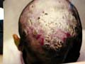
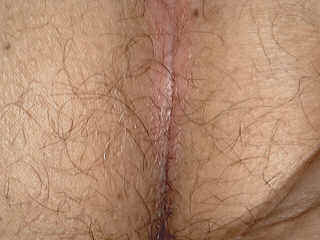
Mucous membranes: the tongue, anogenital area may be involved by psoriasis in the form of whitish patches.

Nails may be involved showing transverse ridges or pitting of the nail plate.

Scalp lesions may extend beyond the hairline and this usually differentiates psoriasis from seborrheic dermatitis, which have greasy scales.

Psoriatic arthropathy: arthropathy is rare and occurs in chronic cases. Psoriasis may be found concomitant with other skin diseases such as lichen planus, vitilligo, lupus erythematosus pemphigus and pemhygoid.

Psoriasis in children

Psoriasis is quite common in children, although congenital psoriasis is very rare. The disease appears first in the scalp, where lesions appear as scaly patches on the scalp and may spread later to involve different skin sites mainly on the extremities and trunk.

Napkin psoriasis:

Flexural and guttate psoriasis is most common in children. Apart from the common forms, several other patterns of psoriasis occur in childhood. The disease often first appears on the scalp.

Flexural and psoriatic intertrigo in children: Interdigital Tinea is uncommon in children and a toe-cleft intertrigo may be psoriatic. Other flexural forms also occur.

Infantile and juvenile pustular psoriasis

Although this type can affect any age in childhood where in some cases the onset may begin in the first year. The lesion is usually circinate or annular. Systemic symptoms are often absent and spontaneous remissions occur.
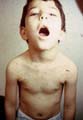
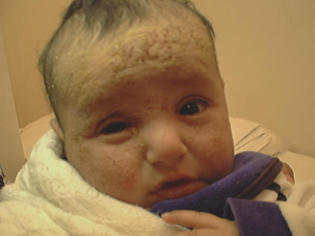
                                                            Fig. 269b. Infantile psoriasis

                                                    ( Mis-diagnosed as seborrheic dermatitis)


A history of an eruption diagnosed as seborrheic dermatitis, napkin dermatitis or napkin psoriasis is obtained. Fever and toxicity may accompany more severe forms.

The majority of children are aged 2 to 10 years old at the time of onset.


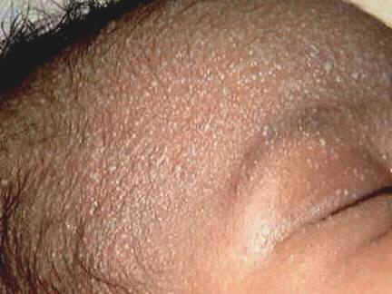
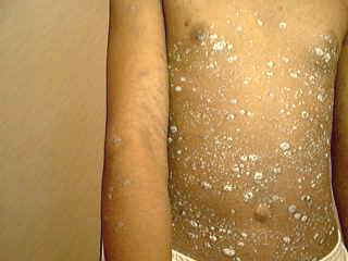
                                                                                           Fig. 269 b. Guttate psoriasis


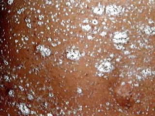
                                                               Fig. 269 c. Rupid psoriasis


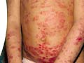
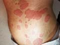
Guttate psoriasis

Small lesions, appearing more or less generally over the body, particularly in children and young adults, usually after acute streptococcal infections. The lesions appear as small rounded or oval patches on the trunk, limbs, scalp and face.

Rupid or unstable type

This type of psoriasis is characterized by scaly hyperkeratotic lesions with concave surfaces, which are unstable and may proceed to pustular, or erythrodermic type.

Intensive systemic or topical steroid therapy, hypocalcaemia, acute infection, over treatment with tar, Dithranol or PUV irradiation and perhaps severe emotional upset may precipitate this condition.

Erythrodermic psoriasis

This type of psoriasis is usually a manifestation of exacerbation of pre-existing psoriatic lesion as the unstable type. This may follow sensitivity reaction to different topical applications as tars, anthralin, PUVA, infections, hypocalcaemia, systemic or topical steroids as colbetasol used on an extensive body surface for a long period.


Fig. 270. Erythrodermic psoriasis


             Fig.270b. Erythrodermic psoriasis


The characteristics of the disease are often lost, the whole skin is involved and there is severe itching (in contrast to other types of psoriasis, where skin lesions are usually non-itchy). The patient is febrile and ill. The course is often prolonged where relapses are frequent and may be fatal.


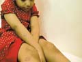
                                                           Fig. 274a. Flexural psoriasis


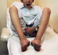
Follicular psoriasis: occurs on the extensor prominence of elbows and knees.

Pustular psoriasis of Zumbach

This type of psoriasis is severe, generalized and may be fatal. This is considered a severe type of psoriasis, due to extensive skin involvement and usually is accompanied by systemic manifestation such as hepatitis.


Fig. 271. Pustular psoriasis


Fig. 272. Erythrodermic & Pustular psoriasis

Clinical Features

The onset is sudden, where iodides and salicylates may act as a triggering factor. Pus is formed periungual followed by generalized erythema.


Fig. 273. Follicular psoriasis


Fig. 274. Follicular psoriasis

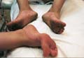
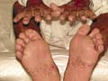
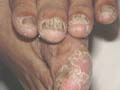
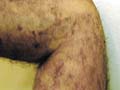
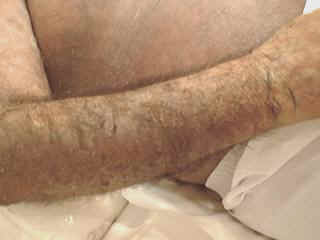
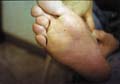
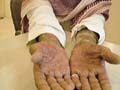
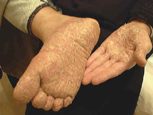
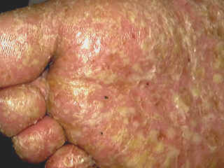
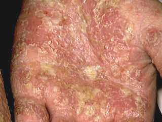
Skin Manifestations

The main symptoms are pruritus, burning of the skin besides fever; fetid odor develops due to extensive exfoliation, and oozing. Yellowish dry crust is formed over a reddish brown shiny surface after drying of the lesion.

Annular and other lesions may be seen in acute generalized pustular psoriasis but are more characteristics of the sub acute or chronic forms of widespread pustular psoriasis. Lesions begin as discrete areas of erythema, which become raised and edematous.

Systemic steroid therapy carries the hazard of disseminated secondary infections with varicella and other viruses.

Mucous membrane lesions of the lips and tongue may lead to superficial ulceration and scaling.

The prognosis is variable but the disease may terminate spontaneously or develop into more severe manifestations.

Generalized pustular psoriasis is well documented in childhood, while arthropathic psoriasis is rare in children.

Histopathology of pustular psoriasis: there is characteristic spongiform pustules in the upper epidermis lined with swollen epidermal cells and contain polymorph nuclear leukocytes.

Treatment of pustular psoriasis: this type of psoriasis is treated with systemic steroids and ACTH.

Psoriatic blepharitis and angular stomatitis

The disease may mimic chronic blepharitis or perleche, usually unilaterally, with a small plaque of psoriasis on one eyelid extending to the lid margin or on the cheek at the angle of the mouth.

Psoriasis of hands and feet: More extensive chronic lesions may occur with persistent dryness, hyperkeratosis and fissuring. Pitting of the fingernails may be the only manifestation for months or even years.

Psoriasis is usually less severe in summer and worse in winter and this may be attributed to the beneficial effect of ultraviolet light of the sun. This phenomenon is clear in cold areas, where sun disappears for longer time in winter than in the tropical areas.

Acrodermatitis continua of Hallopeau

Pustular psoriasis is a disease of middle life. Acrodermatitis may be seen in children.

The first lesion starts on a finger or a toe, related usually to a minor trauma or infection. The skin over the distal phalanx becomes red scaly and pustules develop.

The nail folds and nail bed may be involved leading to nail dystrophy. The proximal edge of the lesion is bordered by a fringe of undermined epidermis, irregular, often soddens and sometimes proceeded by a line of vesiculo-pustules. The nail plate may be completely destroyed.

Bony changes can occur with osteolysis of the tuft of the distal phalanx. The free end of the digit may become wasted and tapered, mimicking scleroderma. In such digits, the circulation may be secondarily affected so that discomfort is greatest in cold weather.

Acute Palmoplanter Pustular Psoriasis (Pustular bacterid)

This term was first used to describe a rare, acute, monomorphic eruption of sterile pustules occurring on all aspects of the hands and feet. It begins abruptly so that within a few days large numbers of small 2-4 mm pustules are distributed on the palms, soles, and palmoplanter aspects of the digits. Sometimes lesions are seen on the dorsa of the hands and feet.


                                                    a) 

                                                                  Fig. 274a,b,c Pustular Psoriasis

                                                                b)   

                                                                c)   

The eruption has a tendency to settle in a few weeks and sometimes only one crop of pustules develops.

Differential Diagnosis of Psoriasis

Psoriasis may simulate different skin diseases

Seborrheic dermatitis

Sometimes it is not easily to differentiate seborrheic dermatitis from psoriasis.


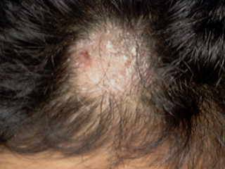
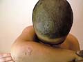
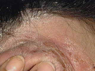
                                                                                    Fig. 274b. Psoriasis of the scalp and the skin

                                                                                                      (Silvery and dry scales)


                                                                   Fig. 274c. Seborrheic dermatitis(Greasy and fine scales, for D.D )

In seborrheic dermatitis the lesions are lighter in color, less well defined and covered with a dull or branny greasy scales.

Eczema at times develops a psoriasiform appearance, especially on the legs. Hyperkeratotic eczema of the palms is a common cause of misdiagnosis.

Lichen planus

The violaceous color, glistening surface and presence of oral changes are usually decisive.

Lichen simplex can resemble psoriasis closely, particularly on the scalp and near the elbow. The intensified skin markings, rather ill defined edge and the marked itching are characteristic.

Pityriasis lichenoides chronica

Can closely resemble guttate psoriasis but the lesions are usually less evenly scattered and have a brownish-red or orange-brown color and are capped by an opaque, soft, ‘mica-like‘ scale.

Candidiasis

Candida lesion presents with a glistening, deep red color suggestive of psoriasis, particularly in the flexures, but scaling tends to be confined to the edge with small satellite pustules and papules which are usually evident outside the main area.

Tinea cruris

Has a well-defined, often polycyclic edge, but Trichophyton rubrum infections, especially of the palm, cause difficulty in differential diagnosis. If corticosteroids have been applied, scaling may be absent, microscopic examination of the scrapings and culture can settle the diagnosis.

Pityriasis rubra pilaris

May simulate psoriasis. The resemblance of pityriasis rubra pilaris may be close, especially in the erythrodermic phase. The color is generally less distinct and deeply red, follicular lesions that are apparent and the horny thickening has a yellowish tinge.

The psoriasiform lesions of syphilis

May cause difficulty in differentiation. Other manifestations of syphilis as condylomata and other signs besides the serological tests for syphilis help in the differential diagnosis.

Other skin diseases

Porokeratosis of Mibelli on the palms and soles, patches of Bowen‘s and Paget‘s disease and penile erythroplasia may resemble psoriasis, but the lesions are usually solitary except in chronic arsenical poisoning. A biopsy may be necessary. 

Drug eruption

This must be distinguished from psoriasis, particularly the reaction induced by the beta-blocker (practolol).

Parakeratosis pustulosa

Is an eczematous eruption seen in young children and commonly mistaken for psoriasis, atopic dermatitis or tinea. It affects the skin around one or more fingernails or toenails, causing subungual hyperkeratosis and thickening of the free edges of the nail. Scaling is more marked than pustulation and the lesions have a chronic course.

Treatment of Psoriasis

The physician can treat mild cases of psoriasis. Referral to a dermatologist may be necessary especially in the following conditions:

Wide spread and disseminated lesions.

Exfoliative lesions and erythrodermic reactions.

Pustular psoriasis.

Lesions not responding to the traditional types of medications.

Recurrent lesions.

  1. General and non-specific measures.

    Rest and mild sedation.

    Removal from a troublesome environment, a holiday or a short stay in hospital may all help.

    Relaxation in an area where high sunlight exposure is possible, such as the Dead Sea coast.

    Patient reassurance is very important, convincing him or other contactants that this type of skin disease is not contagious, can be treated and needs some patience.

    Much care should be considered in treating psoriasis of infants and young children, where medications used for adults may cause serious side effects for these age groups.

  1. Tar therapy

    Tar has been used as topical therapy for more than a century. Goeckerman popularized its use in psoriasis. Daily application of 2-5% crude tar, combined with a tar bath and ultraviolet light, has been used.

    Scalp lesions we use an oily preparation of tar that is the oil of cade.

    Many commercial creams, lotions, ointments, gels and shampoos containing tar extracts are available, which often partially control some cases of psoriasis but are disappointing in severe disease.

    Tar alone is certainly active in psoriasis as is UVB alone. Coal tar seems to sensitize the skin to UVA but not to UVB and phototoxicity is of photodynamic type.

    Nevertheless UVB is more valuable than UVA in conjunction with tar and probably UVB erythema thresholds prevent UVA exposure sufficient to cause photosensitization in the Goeckerman regime.

    A combination of 5% crude coal tar and Dithranol was found to be as effective.

    Primary irritation is uncommon except in unstable psoriasis, and on the face, genitalia and in the flexures. Allergic contact dermatitis does occur, but is rare.

    Folliculitis is the commonest side effect.

    Reports of carcinoma in the site of local coal tar treatment is few but may occur.

    Contraindications of Tar Therapy.

    Infants and young children.

    Anogenital area and axillary folds.

    Erythrodermic or generalized pustular psoriasis.

    Pre-existing folliculitis.

    Severe acne.

    Sensitivity to tar and its derivatives.

  1. Topical steroids

    It is of prime importance to begin treatment with mild topical applications especially in children. If too vigorous successions of therapeutic procedures are applied, it may be soon found that all effective methods of treatment have been exhausted.

    Mild cases may need only simple emollients or mild steroid topically as hydrocortisone ointment.

    Combination of the steroid ointment with salicylic acid or tar, although it can give good results in older age groups, in children much care should be kept in mind when prescribing such combinations.

    Superior results with topical steroids occluded by a hydrocolloid dressing as opposed to plastic film or using (Cordran tape) which is special tape with fluorinated steroid. This method has also its drawbacks. Apart from the cutaneous adverse effects, the most potent preparations or high doses easily suppress plasma cortisol levels, especially when used for a long period on a wide area of the skin. This may lead to more absorption of the steroid and more serious side effects and may induce pustular psoriasis.

    N.B.

    An amount of 7-g daily clobetasol propionate 0.05% or 0.05% Betamethasone dipropionate was sufficient to suppress morning plasma cortisol levels in 20% of patients.

    Scalp lesions need mild steroid lotion and tar shampoos.

    Psoriasis of face and flexural

    Much care should be taken when treating lesions of face, flexural and genitalia, using only mild steroid, where potent steroids can cause more complications locally for the delicate skin of such areas.

  1. Vitamin D3 analogues: Calciptrol (Daivonex) applied once daily to the lesions may give good results especially, when mild steroid combined with salicylic acid and are used twice daily.
     

  2. Psoralenes with PUVA or sun light exposurev

    Care also should be considered in using PUVA, PUVB as a line of treatment due to unwanted side effects and exacerbation of lesions in some patients where such medications are not indicated for young age groups less than 12 years of age. Psoralene tablets are taken two hours before the lesions are exposed to PUVA. The dose of Psoralenes are adjusted according the weight of patients.

    Protect the eyes by special glasses from the effects of PUVA. If PUVA can‘t be used, exposure to sunlight which should be in the morning and before 3:00 p.m., where ultraviolet rays decrease after that.

  1. Methotroxates:

    These drugs are not used for infants and children due to their hazardous effect in the young age groups. In adults and older age groups, these may be used for reluctant, severe psoriasis, which is not responding to all traditional treatment. Methotroxates should be used under strict observation and after thorough investigations especially blood count and liver functions.

  1. Other medications used for psoriasisis:

    Clofazimine, Dapsone and Sulphapyridine are known to enhance neutrophil phagocytosis and may be helpful in pustular psoriasis.

    Infliximab (3 mg/kg) -(5 mg/kg) is a monoclonal antibody that specifically binds to tumor necrosis factor- , blocking its biologic activity. Infliximab 3-5 mg/kgm. Is well tolerated and  can cause  significant improvement  of psoriasis


  1. Corticosteroids

    Corticosteroids should be used with extreme caution. Very high doses of prednisolone followed by an abrupt withdrawal in the treatment of acrodermatitis continua has precipitated generalized pustular psoriasis.

    Small doses of triamcinolone, in dosage not exceeding 6 mg daily initially and with maintenance doses of 2-4 mg daily may be effective especially in severe and erythrodermic lesions.

  1. Retinoids

    Vitamin A has long been recognized to have profound effects on epithelial differentiation and the toxicity of hypervitaminosis A is well known.

    Deficiency causes cutaneous hyperkeratosis and squamous metaplasia of mucous membranes.

    The term ‘retinoid‘ has been applied to a family of natural and synthetic analogues of vitamin A.

    a. Isotretinoin

    Isotretinoin was reported to improve generalized pustular psoriasis. Isotretinoin was also found to be less effective than etretinate in the treatment of chronic plaque psoriasis.

    b. Etretinate.

    Etretinate has been shown to induce remission in pustular types and appears to be significantly more effective than PUVA.

    In an attempt to minimize the side-effects associated with long-term, high dose treatment, clinical improvement was induced with high doses (70 mg etretinate adult dose daily) followed by maintenance of remission with lower doses (30 mg daily).

    c- Acetrtin ( Soriatane) : This is a rettenoid oral drug newly FDA approved for severe types of psoriasis.It should be taken into consideration all the precautions and contraindications of its use mainly in child bearing age groups.

  1. Cyclosporin (1-6 mg/kg body weight/day) was found to improve pustular lesions in adults. Withdrawal of the drug lead to rapid relapse. The side effects of Cyclosporin need precautions during treatment.
     

  2. Zidovudine (azidothymidine):

    AIDS-associated psoriasis has been reported to clear with oral Zidovudine. This drug may be the treatment of choice for retinoid-resistant, AIDS-associated psoriasis, as agents such as methotrexate, Cyclosporin, PUVA and possibly even topical steroids may be contra-indicated.

  1. Hydroxyurea

    Compared with methotrexate has less side effects as anorexia, nausea and hepatotoxicity.

    Dosage should rarely exceed 0.5 g thrice daily and sometimes 0.5 g once or twice daily suffices for maintenance therapy.

  1. Fish oil

    The mechanism of action may involve interference with arachidonic acid metabolism which is one of the major fatty acid components of fish oil. Fish oil can be used as an adjunctive treatment in psoriasis.

  1. Dead Sea bathing : due to high salt contents may improve some cases especially if bathing is followed by sunbathes.
     

  2. Dialysis and related procedures

    Dialysis has some effect on psoriasis in patients with normal renal function and that peritoneal dialysis is more effective than haemodialysis, possibly because substances of higher molecular weight can be removed in larger quantities.

    16-Narrow band treatment of psoriasis

    Narrow-band UVB Photography

    Narrow band laser treatment for psoriasis and vitilligo has been considered recently as an effective line of therapy for psoriasis especially cases which don't respond to the traditional methods of treatment.Although this type of treatment is expensive,yet it is the line of choice for those who can afford paying for narrow band costs.

    Narrow-band UVB refers to a specific wavelength of ultraviolet (UV) radiation, 311 to 312 nm. This range has proved to be the most beneficial component of natural sunlight for Psoriasis and is promising in the treatment of some other skin conditions including atopic eczema and vitiligo. 

    Compared with broadband UVB, in the treatment of psoriasis, Narrow band UVB treatment has the following features:
    * Exposure times are shorter but of higher intensity. 
    * The course of treatment is shorter
    * It is more likely to clear the psoriasis
    * Longer periods of remission occur before the psoriasis reappears
    For patients with psoriasis severe enough to require phototherapy, narrow-band UVB offers efficacy superior to conventional UVB and appears intrinsically safer than PUVA. However, because it produces more epidermal damage than wideband UVB, the narrowband treatment must be used with careful individualization of dosage and patient monitoring, according to James G. Krueger, MD, PhD.

    17- Alefacept is  a selective immunomodulator drug which is recently approved for the treatment of psoriasis. The drug blocks the effect of the pro-inflammatory cytokine tumor necrosis factor . 


REFERENCES

  1. Iselius L, Williams WR. The mode of inheritance of psoriasis: evidence for a major gene as well as a multifactorial component and its implication for genetic counselling. Hum Genet 1984; 68: 73-6.

  2. Abel EA, DiCicco LM, Orenberg EK et al. Drugs in exacerbation of psoriasis. J Am Acad Dermatol 1986; 15: 1007-22.

  3. Abel EA, Barnes S, Le Vine MJ et al. Psoriasis treatment at the Dead Sea: second international study tour. J Am Acad Dermatol 1988; 19: 362-4 (letter).

  4. Beylot C, Bioulac P, Grupper C et al. Generalised pustular psoriasis in infants and children: report of 27 cases. In: Farber EM, Cox AJ, Jacobs PH, Nall

  5. LM, eds. Psoriasis. Proc 2nd Int Symposium. New York: Yorke Medical Books, 1977: 171-9.

  6. Beylot C, Puissant A, Bioulac P et al. Particular clinical features of psoriasis in infants and children. Acta Derm Venereol 1979; 59 (Suppl. 87): 95-7.

  7. Beckman L, Bergdahl K, Cedergren B et al. Genetic markers in psoriasis. Acta Derm Venereol 1977; 57: 247-51.

  8. Arntzen N, Kavli G, Volden G. Psoriasis provoked by beta-blocking agents. Acta Derm Venereol 1984; 64: 346-8.

  9. Barth JH, Baker H. Generalized pustular psoriasis precipitated by trazodone in the treatment of depression. Br J Dermatol 1986; 115: 629-30.

  10. Baughman R, Sobel R. Psoriasis, stress and strain. Arch Dermatol 1971; 103: 599-605.

  11. Burch PRJ, Rowell NR. Psoriasis: aetiological aspects. Acta Derm Venereol 1965; 45: 366-80.

  12. Belsito DV, Kechijian P. The role of tar in Goeckerman therapy. Arch Dermatol 118: 1982; 319-21.

  13. Bollag W. From vitamin A to retinoids: chemical and pharmacological aspects. In: Orfanos CE, Braun Falco O, Farber EM et al., eds. Retinoids. Advances in Basic Research and Therapy. Berlin: Springer-Verlag, 1981: 5-11.

  14. Bollag W. Chemistry and pharmacology of retinoids. In: Farber EM, Cox AJ, eds. Psoriasis. Proc 3rd Int Symposium. New York: Grune & Stratton, 1982:175-83.

  15. Corbett M. Controlled trials of PUVA and etretinate for psoriasis. Br J Dermatol 1985; 112: 121-2 (letter).

  16. Dobson RL. The inheritance of psoriasis (editorial). Arch Dermatol 1980; 116: 657.

  17. Danno K, Horio T, Ozaki M et al. Topical 8-methoxypsoralen photochemotherapy of psoriasis. Br J Dermatol 1983; 108: 519-24.

  18. Epstein JH, Farber EM, Nall L et al. Current status of oral PUVA therapy for psoriasis. J Am Acad Dermatol 1979; 1: 106-17.

  19. Krueger GG. Psoriasis; current concepts of its etiology and pathogenesis. In: Dobson RL, Thiers BH, eds. 1981 Yearbook of Dermatology. Chicago: Yearbook Medical Publishers, 1981: 13.

  20. Farber EM, Jacobs AH. Infantile psoriasis. Am J Dis Child 1977; 131: 1266 9.

  21. Farber EM, Nall ML. Epidemiology: Natural history and genetics. In: Roenigk HH, Maibach HI, eds. Psoriasis. New York: Marcel Dekker, 1985: 141-86.

  22. Henseler T, Christophers E. Psoriasis of early and late onset: characterisation of two types of psoriasis vulgaris. J Am Acad Dermatol 1985; 13: 450-6.

  23. Holgate MC. The age-of-onset of psoriasis and the relationship to parental psoriasis. Br J Dermatol 1975; 92: 443-8.

  24. Kavli G, Frde OH, Arnesen E et al. Psoriasis: familial predisposition and environmental factors. Br Med J 1985; 291: 999-1000.

  25. Jefferson J. Napkin psoriasis. Br J Dermatol 1966; 78: 614-15.

  26. Lever WF, Lever GS, eds. Histopathology of the Skin 6th edn. Philadelphia: Lippincott, 1983.

  27. Mahrle G. Meyer-Hamme S, Ippen H. Oral treatment of keratinizing disorders of skin and mucous membraines with etretinate. Arch Dermatol 1982; 118:97-100

  28. Kapp A, Kemper A, Stop E et al. Detection of circulating immune complexes in patients with atopic dermatitis and psoriasis. Acta Derm Venereol 1986; 66: 121- 6.

  29. Lerner MR, Lerner AB. Congenital psoriasis: report of three cases. Arch Dermatol 1972; 105: 598-601.

  30. Pascher F, Wood WS. Erythrodermic psoriasis in children. Arch Dermatol 1956; 74: 173-6.

  31. Wright N. The cell proliferation kinetics in psoriasis. In: Goldsmith LA, ed. Biochemistry and Physiology of Skin. Oxford: Oxford Univer-sity Press, 1983: 203-29.

  32. Zachariae H. Epidemiology and genetics. In: Mier PD, van de Kerkhof PCM, eds. Textbook of Psoriasis. Edinburgh: Churchill Livingstone, 1986: 4-12.

  33. Young E. The external treatment of psoriasis. Br J Dermatol 1970; 82: 510-15.

Top

Contents << Previous Chapter Next Chapter >> Search