|
LICHEN PLANUS
|
|
|
|
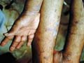
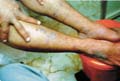
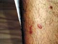
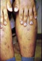
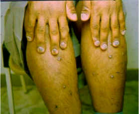
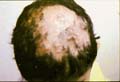
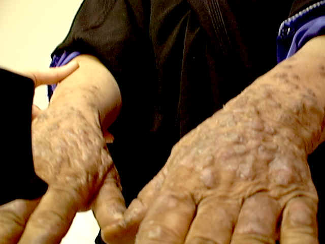
The etiology of lichen planus may be due to immunological factors.There are different data suggesting genetic predisposition. Lichen planus involves the skin and mucous membrane. The primary lesion is violaceous polygonal, flat topped papules which show grayish lines on the surface when examined under a magnifying lens. This is known asWickman‘s stria which is one of the diagnostic criteria for lichen planus. Clinical Varieties Lichen planus has different clinical varieties: Localized type. Lesions of the scalp presents with dry scaly areas, which heal by atrophy and cicatricial alopecia. Lichen planus of the palms and soles may show pigmented, depressed areas besides the primary lesion. Nails may be also involved causing nail dystrophy. Skin lesions may have different shapes or patterns, usually symmetrical in the form of annular, linear or confluent large plaques involving mainly the extremities. Annular type Linear type Follicular lichen planus Guttate lichen planus. Hypertrophic lichen planus affects the lower limbs. Cicatricial type involves mainly the scalp leading to cicatricial alopecia. Lichen tropicus: Lichen planus tropicus is another type of lichen planus involving the sun exposed areas.
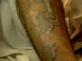
usually in the tropicus. The lesions are characterized by well-defined nummular patches which have a deeply hyperpigmented center surrounded by a striking hypopigmented zone precipitated by excessive exposure to sunlight.
Fig.282b. Lichen actinicus Lichen planus of the mucous membranes: mucous membranes of the oral cavity, bladder, glans penis and rectum may be involved where lesions of the mucous membranes are whitish. Tongue lesions are more on the tip, where the lesions are whitish and the center appear more depressed than the periphery of the lesions. Candida albicans may be associated with lichen planus of the oral cavity. Severe pruritus may accompany the skin lesions, while that of the mucous membranes; the lesions are small, whitish and non-itchy. Lichen planus associated with other diseases. Lichen planus is found to be concomitant with some autoimmune diseases, liver cirrhosis, and other liver abnormalities. So it seems important to screen all patients with lichen planus to investigate such cases thoroughly mainly liver-function tests. Different drugs can induce lichen planus- like reaction. These include beta-blockers as naproxen, quinine, gold, PAS, streptomycin, isoniazid, methyldopa, metropromazine and lithium carbonate. Histopathology Histopathological features of lichen planus are: Thinning of the stratum granulosum. Destruction of the basal layer of saw tooth appearance. Cellular infiltrate mainly lymphocytes below the epidermis. Treatment Antihistamines to relieve the severe pruritus which is usually one of the important distressing features of lichen planus. Topical steroids are not always helpful. Co2 laser may resurface hypertrophic lesions if other methods were tried, as occlusion with potent steroids topically in older age groups. Retinoids: may cause improvement of some cases of lichen planus.
|
||||||||||||
| Contents | << Previous Chapter | Next Chapter >> | Search |