|
DISEASES OF ABNORMAL KERATINISATION
|
|
|
|
Disorder of keratinization is due to a defect in keratin metabolism. In the normal epidermis, as the keratinocytes move from the basal-cell layer to the surface, the process of terminal differentiation and cornification involves complex metabolic changes. Different syndromes, which are related to abnormal keratinization, include:
Ichthyosis is a group of disorders that are characterized by a persistent, non-inflammatory scaling disorder of the skin surface. Types of icthyosis Genetic icthyosis Acquired icthyosis
Two types of icthyosis vulgaris are known; the dominant and the X- linked type.
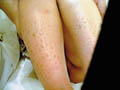
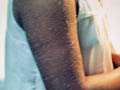
Clinical Manifestations Large scales covering the neck face ears and the flexural surfaces as the axilla, the ante cubital and popliteal areas. The scalp is scaly.
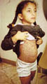
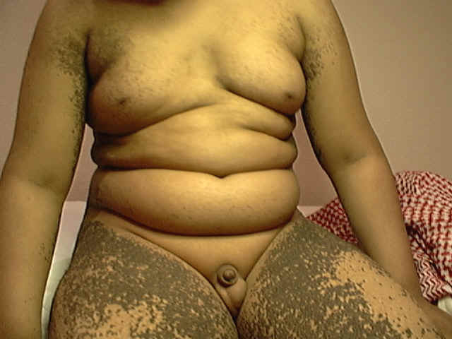
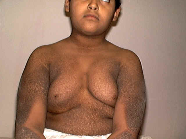
Fig.360b.Icthyosis &KlineFilter's syndrome Histopathology Epidermis: Hypertrophic and hyperkeratotic. Thick granular layer. Treatment Emollients should be used regularly to relieve the dryness and itching and to decrease the tendency to skin fissuring. Preparations containing urea and 2 per cent salicylic acid in a suitable vehicle for severe skin scaling. Care should be taken into consideration when using topical salicylic acid in children due to the possibility of toxic absorption. Some reports showed good improvement of scaling by using 12% ammonium lactate containing lotions. The retinoids group of drugs may give good improvements, but the side effects limit their use in infants and young children and may be reserved for severe reluctant cases in older age groups. Antihistamines may be needed in the presence of itching.
This disease begins usually at birth and is inherited as an autosomal recessive trait. Clinical Features
Histopathology Hyperkeratosis Hyper grannulosis Prominent rete ridges Mild perivascular infiltrate in the upper dermis and mitosis. Treatment
ICTHYOSIFORM
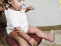
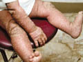
ERYTHRODERMA This is a rare severe type of icthyosis present at birth of unknown etiology and has a high mortality rate. It is believed that lamellar exfoliation of the newborn (collodion baby) is a form of lamellar itchyosis Clinical Features Skin manifestations Infants may be born with a distinctive, tough, inelastic collodion-like membrane covering their bodies. The constricting membrane produces a peculiar position of immobility of the limbs and causes ectropion of the eyelids. The membrane eventually fissures and peels. If the baby survives, the membrane is shed, usually to reveal a more characteristic ichthyosiform abnormality beneath. Uncommonly, normal skin is found beneath the collodion membrane. General Features Affected infants appear very ill, dehydrated and in danger of hypothermia.
This is a more severe form of the collodion baby disorder affecting the skin in utero and causing thick, horny, armor-like plates that cover the entire skin surface. There is usually stillbirth or early death after delivery. This very rare type may have other systemic manifestations. The disease is usually fatal within the first month of life. Clinical Features The skin is dry, hyperkeratotic and fissured. The abnormal inelastic skin, resulting in ectropion and small-deformed ears grossly deforms the facial features. Treatment Some reports show encouraging results with the use of etretinate for the affected infants.
BULLOUS
ICTHYOSFORM ERYTHRODERMA This rare disease is inherited as a dominant gene. Clinical Manifestations The disease appears shortly after birth, and is characterized by generalized erythema in early infancy, hyper keratotic areas, scaling and blister formation. The blisters may occur anywhere, but tend to form at the sites of trauma and as a result may develop over the knees and elbows especially when the child begins crawling and walking. The skin has a thickened, horny, arm-like scales, which are present at birth and shed later on leaving a raw surface. The areas involved are the flexures and the inetertriginous areas. Localized verrucuous lesions and bullous type is known (tichyosis hystrix). This type has to be differentiated from epidermolysis bullosa.
ICTHYOSIFORM BULLOSA OF SIEMEN This is a familial disorder that is present at birth but may be delayed and appears first in the neonatal period or later. Clinical Manifestations Skin manifestations Persistent generalized erythroderma and a fine scaling of the entire skin surface. Uniform hyperkeratosis around the knees, elbows, ankles, the palms and soles. Flexures lesions are characterized by erythema and peeling in localized areas. General manifestations Ectropion and deformed ears are features of more severely affected individuals. Ocular defects, intercurrent infections, mental and growth retardation are common manifestations. Differential Diagnosis In early infancy the disease may simulate staphylococcal impetigo, congenital psoriasis and pityriasis rubra piliaris.
ATYPICAL
ICTHYOSIFORM ERYTHRODERMA This syndrome is characterized by a dry, scaly skin disorder with localized areas of hyperkeratosis, mental retardation, deafness, nail and teeth abnormalities. Treatment There is no specific treatment. Generalized erythema of the affected infant must be carefully nursed. Hypothermia and severe dehydration need special care. Corticosteroids have been administered systemically as a life saving in severe cases. Later in life the use of emollients and keratolytics will be required. Topical retinoic acid may be helpful in some cases. Etretinate, in a dose of 0.5-1.0 mg/kg/day, can provide great relief for some patients, although treatment must be lifelong to maintain improvement.
This is a rare variant of ichthyosiform erythroderma, characterized by: Congenital hemidysplasia. Unilateral Ichthyosiform erythroderma. Limb defects. Some cases show central nervous system abnormalities. Visceral defects. Histologically, there is irregular acanthosis and hyperkeratosis as well as an inflammatory cell infiltrate. Treatment Emollients and keratolytics can help mild cases. Salicylic acid 2-6% in white, soft paraffin can remove hard scaly masses, but care must be taken to ensure that salicylism does not develop. Applications of urea-containing ointment sometimes help, and topical preparations containing hydroxyacids such as tartaric acid, alpha-ketoglutaric acid and pyruvic acid have also been used with some success. Both isotretinoin and etretinate have been found to be of great benefit to severe cases. As with retinoid treatment for other disorders of keratinization treatment has to be lifelong to maintain a therapeutic effect.
This is a rare X-linked dominant multisystem disorder, which is characterized by: The skin disorder often has a distinctive, odd, linear or whorled appearance. Ichthyosiform erythroderma. Follicular atrophoderma. Asymmetrical cataracts. Coarse hair and patchy alopecia. Short stature and short limbs. Frontal bossing and dysmorphic facial features. Radiologically shows calcified stippling of the epiphyses.
This uncommon neuroectodermal genodermatosis appears to be inherited as an autosomal recessive disorder. Clinical Manifestations
ERYTHROKERATODERMA The disorder is a disease of keratinization, starts in childhood and is characterized by large, symmetrical fine scaly plaques of erythema with an orange hue involving the cheeks, shoulder girdle, ankles, wrists and buttocks.
ERYTHROKERATOLYSIS The manifestations appear after birth with dry, hyperkeratotic areas, which exfoliates in sheets spontaneously leaving abraded red skin lesions. The skin is dry and dirty looking due to the hyperkeratosis.
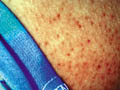
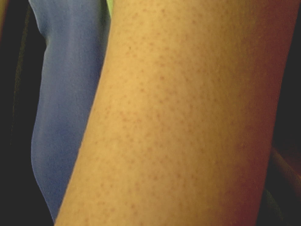
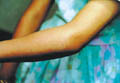
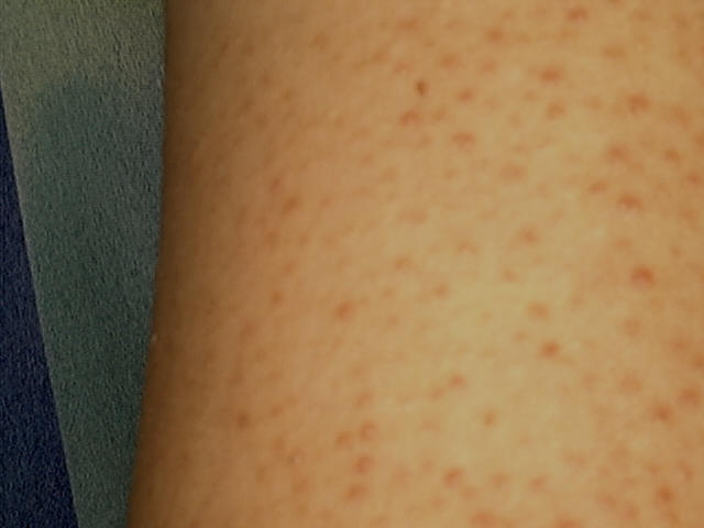
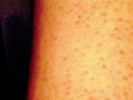
Clinical Picture The lesions appear as small gray to white plugs of keratin that obstruct the mouths of the follicles entrapping the hair. The sites of predilection are the extensor surfaces of the upper arms, thighs and buttocks. Some follicles are completely spared while adjacent ones are grossly plugged showing a long strand of keratin protruding when examined in light (antenna sign).
FOLLICULAR
KERATOSIS Follicular keratosis is a disease of abnormal keratinization that appears in childhood. The skin lesion presents with grouped follicular papules occluded by a projecting keratinous spine. The commonest sites involved are the extensor surface of the extremities, thighs and abdomen.
A number of skin disorders may give rise to similar skin lesions mainly lichen planus and seborrheic dermatitis.
Familial dyskeratotic comedones. This is characterized by scattered comedo-like lesions, inherited as an autosomal dominant condition. The lesions appear around puberty, showing a predilection for the face, trunk, arms, and legs. Retinoid therapy has proved unrewarding.
|
| Contents | << Previous Chapter | Next Chapter >> | Search |