CHAPTER 45

DISEASES OF THE MUCOUS MEMBRANES

Contents << Previous Chapter Next Chapter >> Search

 
Diseases of the mucous membranes are usually more difficult to diagnose than skin diseases. This is partly due to modification of the primary lesion either by the continuous mouth moisture or by different ingredients in the mouth or those reaching the mouth from outside, either through food or mouthwashes or others.

Mucous patches of the mouth should not be mixed with milk patches in babies due to accumulation of milk on the inner sides of the buccal mucosa.

Vesicles or bullae leave a grayish erosion after ulceration, while papular lesions which may be significant on the skin surface become difficult to differentiate in the mucous membranes of the mouth.

Usually mouth lesions are whitish, macerated and soft. Different physiological or pathological factors may affect the buccal and tongue mucosa.

Some of the diseases of mucous membrane, which accompany skin diseases were already discussed  in the previous chapters.

  1. Skin diseases associated with mucous membrane manifestations:

    Pemphigus vulgaris

    Lichen planus

    Lupus erythematosus

    Mucosal candidiasis

    Viral diseases such as Koplik‘s spots of measles and herpes simplex.

    Syphilis


DISEASES OF THE LIPS

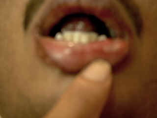
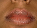
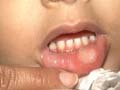
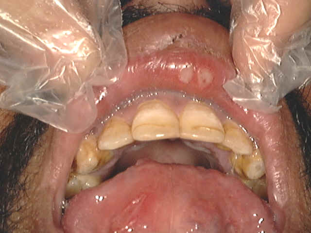
CHEILITIS

Cheilitis is caused by different factors, which may be allergic or inflammatory.

The lower lip is the most common affected.

The lesion may be localized in the lip or may extend to the adjacent mucocutaneous or even to the skin of the face.

Fig. 372 Cheilitis &Glossitis

Etiology of Cheilitis

  1. Infections :

    Candidiasis is the commonest infections that affect babies and young children. The infection may be severe interfering with feeding and may be extensive affecting the whole mucous membranes of the gastro-intestinal tract.

    Bacterial and viral infections are also common.


Fig. 372. Chronic Cheilitis


Fig. 373. Chronic Actinic Cheilitis


Fig. 374. Cheilitis (Contact dermatitis)

  1. Allergic Cheilitis:

    Local hypersensitivity to antibiotics lozenges.

    Mouth washes mainly fluorinated toothpaste.

    Contact dermatitis from certain food stuffs such as lemon, orange, tomato juices and certain food additives.

    In older age groups cosmetics as lip sticks.

  1. Photosensitivity :

    Photosensitivity causes chronic cheilitis. The lips become scaly, fissured and swollen and may lead to leucoplakia that is precancerous.


Fig. 375. Allergic Cheilitis 


Fig. 376. Allergic cheilitis

  1. Cheilitis glandularis: the openings of the mucous ducts become patulous, where mucous drippling mainly during sleeping which on drying causes inflammation of the lip. The mucous glands are enlarged. The condition may be precancerous.


Fig. 377. Actinic chilitis & Leukplakia


Fig.378. Cheilitis(Bacterial infection)

  1. Angular cheilitis. Fissuring of the lips which become painful and bleed. Dental caries may be considered as an important predisposing factor.
     

  2. Cheilitis associated with other diseases and syndromes:

    Leucoplakia affects mainly the lower lips.

    Lichen planus

    Psoriasis

    Lupus erythematosus

    Mongolism

    Vitamin deficiency mainly riboflavin.


                                                                                                                                                                                                  Fig.378b. Chronic ulcerative cheilitis

Drug reaction: Phenolphthalein causes slate blue hyper pigmentation of the lips. Other drugs such as sulfa may cause fixed drug reaction.


Fig. 379. Cheilitis( Drug reaction) 


Fig. 380.  Hairy tongue


Fig.381. Scrotal tongue

GLOSSITIS AND STOMATITIS

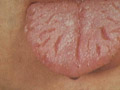
The different factors, which may lead to glossitis and stomatitis include the following:

Malnutrition

Lack of proper hygiene of the mouth

Vitamin deficiency mainly vitamin C and B complex.

Clinical Features

The ulcers are punched out, covered by dirty white pseudomembrane covering the mucous membranes of the tongue, lips, buccal mucosa, tonsils, and the pharynx and may involve the whole respiratory tract mucosa.


                                                                                                                                                                                                                                                               Fig.381b. Glossitis

Herpetic gingivostomatitis

Children and young age groups are commonly infected with herpes virus leading to herpetic lesions of the mouth, tongue and lips. Painful ulcerations are common which may interfere with feeding and increased salivation.

Gangrenous stomatitis

Gangrenous stomatitis occurs in children with poor nutrition, lowered resistance and improper dental hygiene. The lesions manifest with mucosal ulcerations of the mouth and genitalia, which rapidly becomes gangrenous. The condition may extend to the adjacent tissues and may be extensive involving the bones and may be fatal.

Treatment

Proper hygiene of the mouth and correction of the predisposing factors.

Mild mouth wash antiseptics.

Tetracycline suspension orally. This should be kept for few minutes in the mouth to have a local effect.

Care should be taking when treating babies or young children with Tetracyclines. Another oral antibiotic suspension such as Zithromax can be substituted for young age groups.

Systemic antibiotics.


RECCURENT APHTHUS STOMATITIS

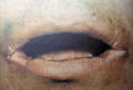
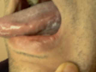
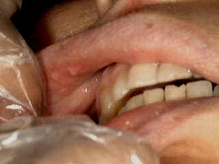
The lesions are recurrent, tender, small shallow erosions surrounded by an erythematous zone affecting the buccal mucosa. These may interfere with feeding and speech. It is believed that streptococcus infection may be the causative of recurrent aphthus stomatitis.


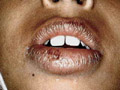
  Fig. 381b. Aphthous stomatitis

The lesions may appear also on the genitalia and has to be differentiated from other diseases of these areas such as viral, fungal and Behcet‘s disease.

Recurrence of lesions may be predisposed by:

Lower body resistance.

Emotional and psychic trauma.

Trauma: self-biting, hard, harsh toothbrushes and other tools used for cleaning of the mouth and teeth.

Hormonal changes.

Allergy to certain types of foods and additives such as tartrazine.

Drug reaction.

Infections such as herpes virus infections may predispose to recurrence.

Differential Diagnosis

It is usually not easy to differentiate such condition from other diseases affecting the buccal mucosa:

Candidiasis

Herpes simplex infections

Vincent‘s angina

Mucous patches of syphilis

Vitamin deficiency mainly scurvy and pellagra.

Behcet‘s disease

Pemphigus


                                                                                                                   Fig.381c. Pemphigus

Lichen planus

Treatment

General measures

Treatment of the predisposing factors.

General hygiene of the mouth and teeth.

Avoid strong mouth antiseptics

Avoid mouthwashes containing allergenic substances.

Avoid contact with certain foods such as citrus, spicy and harsh foodstuffs.

Local treatment

Non-specific medications:

Different local applications were used with variable results.

Tetracycline suspension especially if kept in the mouth few minuets before swallowing, may give good results in some cases.

Xylocaine viscous 2 % (Lidocaine) may be used especially in painful conditions few minuets before feeding for infants and children.

Local topical steroid (Kenalog orabase) or prednisone lozenges may be used if viral or bacterial infections are excluded.

Lesions of the inner sides of lips can be touched gently by swab moistened with salicylic acid 3% and lactic acid 4 % prepared in flexible collodion .


RECURRENT HERPES SIMPLEX OF THE MOUTH

Recurrent herpetic lesions of the mouth are a real problem especially in young age. Herpes simplex virus causes these lesions.

Clinical Features

The lesions appear as small grouped vesicles surrounded by an erythematous zone which later rupture, leaving shallow ulcerations with a red base.


Fig. 382. Herpes Labialis

Diagnosis

Typical clinical picture, which includes painful, grouped vesicles or ulcerations on an erythematous base.

Tzank test: scrapings from the base of the ulcers stained with Wright‘s stain show the multinucleated giant cells.

Immunoflorecent tests are diagnostic.


HERPANGIA

This is a viral disease caused by Coxsackie virus affecting mainly children especially in summer time.

Modes of Infection

It is believed that certain species of flies can transmit the disease.

Direct infection from infected individuals.

Clinical Manifestations

General manifestations

Fever, which may be high, headache, sore throat, anorexia and dysphagia.

Skin manifestations

Skin lesions may appear in different forms mainly:

Exanthematous reaction.

Urticarial lesions.

Pruritic macules.

Mucous membranes manifestations:

Minute papules, vesicles and ulcers surrounded by an erythematous areola appear on the mucous membrane, which are characteristically apparent on the pharynx, tonsils and throat .The lesions appear usually in small groups, which may coalesce and ulcerate.

The ulcers are shallow, grayish yellow and punched.


BEHCET‘S DISEASE
(Aphtosis, occulo-oral genital syndrome)

Behcet‘s syndrome is characterized by:

  • Recurrent ulcerations of the mouth.

  • Genital ulcerations.

  • Eye manifestations such as uveitis and iridocyclitis.

  • Skin manifestations.

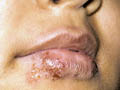
Clinical Features

Skin manifestations

Skin lesions may be:

Erythema nodosum

Erythema multiforme

Acniform eruption

Papulo -pustules, subungual abscess and pyoderma.

General manifestations:

Thrombophlebitis

Arthritis: polyarthritis.

Cerebellar syndrome resembling multiple sclerosis.

Mucous membrane manifestations

Mucous membrane lesions are single or multiple with dirty grayish base surrounded by red halo. Mucous membrane of the buccal mucosa, palate, lips and tongue may be involved. Ulcerations may leave scarring

The ulcers are tender and cause severe pain that may interfere with eating, speech and gives a foul smell of the mouth.

Genital manifestations

Ulceration of the scrotum, penis and urethra.

Ulceration of labia, vagina and cervix.

Ulceration of the anal area, crural areas and perineum. The ulcers are extremely painful.

Healing of ulcers may lead to scarring and deformities.

Eye manifestations

Severe peri-orbital pain and photophobia.

Conjunctivitis, hypopyon, iritis, iridocyclitis.

Blindness may be the end stage due to optic nerve atrophy.


REITER‘S DISEASE

This syndrome occurs chiefly in young males and may affect children.

Clinical Features

This syndrome is characterized by triad manifestations:

Eye: Conjunctivitis, iritis, and keratitis.

Urethritis: Non-bacterial, painful and bloody urination and pyuria. Cystitis, prostatitis and seminal vesiculitis are common

Arthritis: Swollen and tender joints.

General manifestations: Fever, weakness and weight loss.

Skin manifestations: Multiple small, yellowish vesicles and ulcers on the palms and soles. Lesions of the palms and soles become thickly hyperkeratotic and crusted resembling keratoderma blenorrhagica.

Mucous membrane manifestations: Mucous membrane lesions are superficial ulceration of the genitalia and mucous membranes of pharynx and hard palate.

Cardiac manifestations: are rare manifestations of the disease. Endocarditis, myocarditis, pericarditis and aortic insufficiency are uncommon manifestations.


DRYNESS OF THE MOUTH

Dryness of the mouth may be due to different local or systemic factors.

Etiology

  • Drugs: anticholinergic or sympathomimetic activity is the most common cause of dryness of the mouth. Atropine, belladonna, panthene, dimenhydrinate (Drammamine), antidepressants, Phenothiazines and antihistamines.

  • Diabetes.

  • Mickulicz’s syndrome.

  • Sjogren‘s syndrome.

  • Xerostomia.

  • Psychogenic factor.

  • Sarcoidosis.

  • Severe dehydration.


ORAL PIGMENTATION

Etiology

  • Race is the most important cause of oral pigmentation.

  • Addison‘s disease.

  • Melanoma.

  • Laugier-Hunziker syndrome.

  • Pigmentary incontinence

    Peutz-Jegher‘s disease is the association of circumoral and sometimes intra oral melanosis with small intestinal polyposis.

  • Smoking may cause a grayish-brown oral hyperpigmentation.

  • Gold salts may cause purplish gingival discoloration.

  • Lichen planus: may cause oral white patches.

  • Drugs: antimalarial drugs such as chloroquine, quinacrine and hydroxychloroquine drugs may cause hyperpigmentation of the mucous membranes.

Mepacrine: causes yellow discoloration of the buccal mucous membranes.

Amodiaquine or quinidine causes blue-black pigmentation.

ACTH therapy: may cause brown pigmentation.

Busulphan, causes blue line of gums


Fig. 383. Oral Hyperpigmentation

Cytotoxic drugs.

Phenothiazines.

Anticonvulsants.

Chloropromazine: may cause purple or yellowish brown pigmentation of the skin and mucous membranes.

Minocycline: may produce blue-gray gingival pigmentation.

Heavy metals.


PEUTZ-JEGHER‘S SYNDROME

This syndrome is characterized by pigmentation and intestinal polyposis.

The pigmented macules may be present at birth and usually appear in infancy and early childhood, but may develop later in life. The oral mucous membrane is almost constantly involved. The oral pigmentation is usually permanent, but the macules on the lips and skin may fade after puberty. Rarely, the nails may be pigmented, diffusely or in longitudinal bands.

Clinical Features

Mucous membrane manifestations

Round, oval or irregular patches of brown or almost black pigmentation 1-5 mm in diameter are irregularly distributed over the buccal mucosa, the gums, the hard palate and the lips, especially the lower. Mucosal and facial pigmentation without evidence of intestinal polyposis may be found in relatives. Intestinal polyposis may cause repeated bouts of abdominal colic, vomiting, intussusceptions, malignancy and rectal bleeding.

Skin manifestations

The pigmented macules on the face are smaller, dark often under 1 mm  and are concentrated around the nose and mouth.

Larger macules may be present on the hands, feet, palms and soles.

Systemic manifestations

Abdominal pain is usually caused by intestinal obstruction.

Rectal bleeding is common and haematemesis may occur with gastric or duodenal polyps.

Anemia.

Differential Diagnosis

Addison‘s disease

Skin freckling

Gardner‘s syndrome


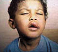
WHITE PATCHES OF THE BUCCAL MUCOSA

Different factors may cause white patches of the buccal mucosa.

Etiology:

Infections: of the buccal mucosa such as herpetic lesions may cause white patches surrounded by an erythematous area.

Thrush: produces oral white patches.

Mucous patches of syphilis: Grayish white painful patches, slightly elevated and surrounded by an erythematous area. The buccal lesions occur most frequently on the inner lower lips, tongue and other parts of the mouth. The mucous patches are highly contagious and serological tests of syphilis are positive.

Lichen planus.

Pemphigus

Benign mucosal pemphigoid

Hairy leucoplakia: is a white lesion that appears on the tongue and whitish flaky pseudomembrane on an erythematous base.

Culture will reveal the causative organism.

Leucoplakia: lichenoides lesions are associated with various drugs, liver disease or graft-versus-host disease.

White sponge nevus: white, thickened spongy patches appear on the inner sides of lips, cheeks and sides of the tongue

Carcinoma may present as a white lesion.

Dyskeratosis congenita causes white patches


REDNESS OF THE BUCCAL MUCOSA

Etiology

  • Chronic marginal gingivitis is the usual cause of gingival redness.

  • Herpes simplex stomatitis.

  • Pellagra: The sides of the tongue are erythematous and soggy which may become later beefy-red.

  • Mal absorption: The mucous membrane of the tongue and buccal cavity is red.

  • Lichen planus.

  • Mucous membrane pemphigoid.

  • Allergic conditions of the buccal mucosa due to mouth washes, chewing gums, toothpaste and others.

  • Erythroplasia.

  • Haemangiomas or neoplasms such as carcinoma.

  • Wegener‘s granulomatosis.

  • Kaposi‘s sarcoma.

  • Telangiectasia: may be a manifestation of hereditary hemorrhagic telangiectasia or systemic sclerosis or may follow radiotherapy.

  • Candidiasis

  • Lichen planus

  • Lupus erythematosus.

  • Kaposi‘s sarcoma may present as a red, purple, brown or bluish macule or nodule.

  • Iron, folate or vitamin B12 defficiency may produce the red tongue.

  • Geographical tongue may also present with red patches.


HALITOSIS

Cacogeusia (an unpleasant taste in the mouth) and halitosis are caused by different factors mainly:

Etiology

  • Poor oral hygiene.

  • Oral or nasal infections.

  • Starvation.

  • Foods

  • Fasting

  • Drugs

  • Psychogenic disorders

  • Systemic disorders such as suppurative respiratory infections, hepatic or renal failure, diabetic ketosis or gastrointestinal disease.


LOSS OF SENSE OF TASTE

Etiology

  • Lingual nerve trauma.

  • Xerostomia

  • Drugs such as penicillamine.

  • Psychotic disorders.

  • Neurological disease such as cerebral metastases, and lesions affecting the chorda tympani may be responsible for loss of the sense of taste.

  • Anosmia commonly produces an apparent loss of the sense of taste.


REFERENCES

  1. Abraham-Inpijn L. Oral and otal manifestations as the primary symptoms in Wegener‘s granulomatosis. J Head Neck Pathol 1983; 2: 20-2.

  2. Basu MK, Asquith P. Oral manifestations of inflammatory bowel disease. Clin Gastroenterol 1980; 9 (2): 307.

  3. Browning S, Hislop S, Scully C et al. The association between burning mouth syndrome and psychosocial disorders. Oral Surg 1987; 64: 171-4.

  4. Budzt-Jorgensen E. Oral mucosal lesions associated with the wearing of removable dentures. J Oral Pathol 1981; 10: 65-80.

  5. Berger RS, Mandel EB, Hayes TJ et al. Minocycline staining of the oral cavity. J Am Acad Dermatol 1989; 21: 1300-1.

  6. Cotaldo E. Solar cheilitis. J Dermatol Surg Oncol 1981; 7: 289-95.

  7. Dorey JL, Blasberg B, MacEntee MI et al. Oral mucosal disorders in denture wearers. J Pros Dent 1985; 53: 210-13.

  8. Hartman KS. Histiocytosis X. A review of 114 cases with oral involvement. Oral Surg 1980; 49: 38-54.

  9. Hietanen J. Clinical and cytological features of oral pemphigus. Acta Odontol Scand 1982; 40: 403-14.

  10. Laskaris GC, Sklavounou A, Stratigos J. Bullous pemphigoid, cicatricial pemphigoid and pemphigus vulgaris: a comparative clinical survey of 287 cases. Oral Surg 1982; 54: 656-62.

  11. Lamey PJ, Lewis MAO, Rees TD et al. Sensitivity reaction to the cinnamaldehyde component of toothpaste. Br Dent J 1990; 168: 115-18.

  12. James J, Ferguson MM, Forsythia A et al. Oral lichenoid reactions related to mercury sensitivity. Br J Oral Maxillofac Surg 1987; 25: 474-80.

  13. Kerr DA, McClarchey KD, Regezi JA. Allergic gingivostomatitis (due to gum chewing). J Periodontol 1971; 42; 709-12.

  14. Lamey PJ, Carmichael F, Scully C. Oral pigmentation, Addison‘s disease and results of screening. Br Dent J 1985; 158: 297-305.

  15. Marks R, Radden BG. Geographic tongue: a clinicopathological review. Aust J Dermatol 1981; 22: 75-9.

  16. Manton S, Scully C. Mucous membrane pemphigoid. Oral Surg 1988; 66: 37-40.

  17. Nisengard RJ, Nieders M. Desquamative lesions of the gingiva. J Periodontol 1981; 52: 500-10.

  18. Picascia DD, Robinson JK. et al. Actinic cheilitis, a review of the aetiology, differential diagnosis and treatment. J Am Acad Dermatol 1987; 17: 255-64.

  19. Schoenfeld RJ, Schoenfeld FI. Angular cheilitis. Cutis 1977; 19: 213-16.

  20. Scully C. Chronic atrophic candidosis (leading article). Lancet 1986; ii: 437-8.

  21. Scully C. Orofacial manifestations in chronic granulomatous disease of childhood. Oral Surg 1981; 51: 148-51.

  22. Scully C, Porter SR. The mouth and skin. In: Verbov JL, ed. New Clinical Applications in Dermatology: Relationships in Dermatology Vol 8. Lancaster: MTP Press, 1988: 1-34.

  23. Touyz LZG, Peters E. Candidal infection of the tongue with non-specific inflammation of the palate. Oral Surg 1987; 63: 304-8.

  24. Theaker JM, Porter SR, Fleming KA. Oral epithelial dysplasia in vitamin B12 deficiency. Oral Surg 1989; 67: 81-3.

  25. Walker DM, Stafford DG, Huggett R et al. The treatment of denture-induced stomatitis. Br Dent J 1981; 151: 416-19.

  26. Winer LH. Black hairy tongue. Arch Derm 1958; 77: 97-103.

Top

Contents << Previous Chapter Next Chapter >> Search