|
Viruses have different structures, compositions, pathological and clinical
manifestations. Viruses are extremely small in size that
are capable of passing the bacterial filtrate.
General
considerations
Small viruses lack
enzyme system while large ones such as the organisms of trachoma, lymphgranuloma venerum have
some and this is why the latter group is susceptible to chemotherapy, while
the first is not.
Viruses are obligate
intracellular parasites, the principle site of attack in the skin is the epidermis . These may cause localized or systemic
manifestations that varies from erythematous
lesions, vesiculation, ulceration
, scarring or severe constitutional symptoms .
HERPES
SIMPLEX
Herpes simplex is the
most common of all viral infections. The herpes virus causes infection.
Many patients become carriers.
Herpes infection is a
contagious disease and spread by droplet infection, contact as in kissing
or contact with lesions of infected individuals and infected fomites .
Clinical picture
of herpes simplex
The incubation period
of herpes infection is from 4-5 days . The lesions
may be cutaneous or mucocutaneous.
Primary lesions may affect any age but is most common in children
, while new born under 4 month of age has transferred maternal
antibodies and are rarely infected .
In the majority of
cases infection is subclinical or asymptomatic .
Primary and recurrent
infections are highly infectious and heal completely but the virus can be
detected in the cells for many years.
Tingling and burning
sensation appears at the involved site, then few
small grouped vesicles on an erythematous base
appear which rupture and heal within two days. The course may be longer
when secondary bacterial infection complicate the
lesions.
Herpes infection is
characterized by an acute eruption of grouped vesicles upon an erythematous base most frequently on the mucocutaneous junction. The symptoms may be very mild
attacks or very severe even fatal in newborn.
Infection may be
primary in individuals who have no specific neutralizing antibodies or recurrent
which is exceedingly common in individuals who posses specific antibodies.
|

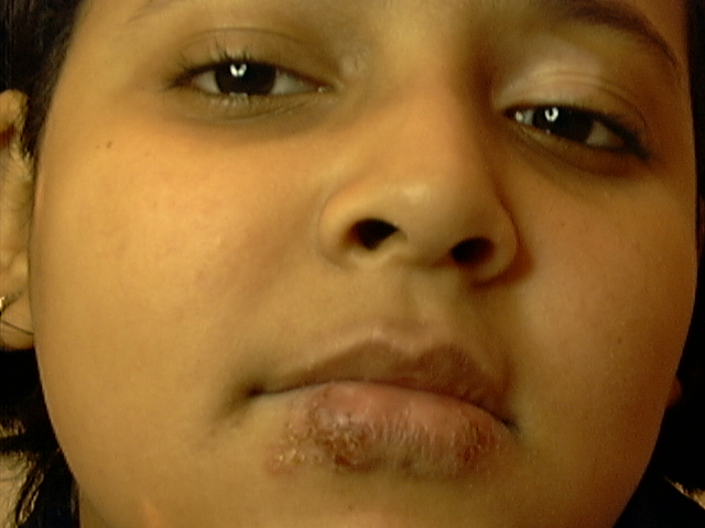
Fig. 87. Herpes labialis
|
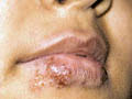
Fig. 88. Herpes labialis
|
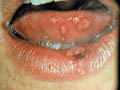
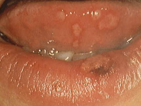
Fig. 89. Herpes of lip
& Tongue
|
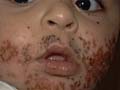
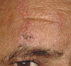
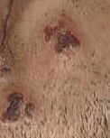
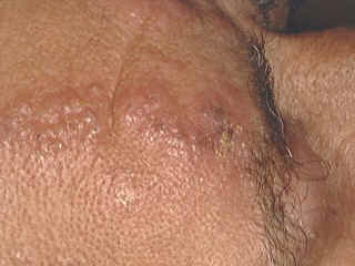
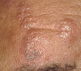
Fig. 90-. Eczema herpeticum
|
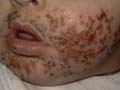
Fig. 91. Eczema herpeticum
|
NEONATAL
HERPES SIMPLEX
Herpes simplex virus (HSV)
infection in the newborn is generally a serious disease with a high
mortality rate.
Modes of infection
Transmission of the
HSV type II by contact with infected genital tract secretions during
delivery.
Intra-uterine HSV infection may occur, due both to transmission across the
placenta, or to an ascending from the infected genitalia if the mother has
prolonged rupture of the fetal membranes.
Contact with
non-genital sites, both maternal and non-maternal.
Clinical features
The skin lesions are isolated or grouped
vesicles that appear mainly on the scalp and face. Occasionally generalized
bullous lesions or widespread erosions may occur
without obvious vesicles . Healed lesions may show
atrophy or scarring simulating epidermolysis bullosa .
When the infection is
acquired during birth, the initial lesions have a predilection for the
scalp in vertex presentations, and the perianal
area in breech presentations
Oral lesions are also frequent, and take
the form of erosions on the tongue, palate, gingiva
and buccal mucosa.
Central nervous
system
involvement
Fatal cases may occur
when infection is disseminated, even when appropriate antiviral therapy is
given.
Early recognition and
adequate early treatment with Acyclovir does appear to protect infants from
dissemination of infection where this is initially confined to the skin .
Types of herpes
simplex
Type 1: causes cutaneous and oral lesions.
Some rare genital lesions are due to this type.
Type 2: this is the cause of herpes progenitalis.
Different clinical
types of Herpes Simplex
1.
Primary herpes simplex infection
Cutaneous lesions appear as painful grouped vesicles on an erythematous base around the mouth that ulcerate
leaving a painful ulcer.


Fig. 91a. Primary herpes simplex
Different clinical
variants of herpetic lesions may appear with different clinical pictures
according to the site involved either in the skin or the mucous membrane.
2. Mucous membrane
lesions
This is a very common
viral infection in young children between the age of 2-5 years
, in older children and young adults . The condition begins with
fever and the sudden development of painful oral lesions, which ulcerate.
These may be misdiagnosed as Vincent‘s angina, aphthous
stomatitis or other ulcerating bullous diseases. The mucous membrane becomes red,
swollen and painful with ulceration. These are considered very important
cardinal signs of herpetic infection of the mucous membranes
.
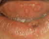
Fig.91b. Herpes of the mucous
membrane
Extensive involvement
of the mucous membrane of the mouth, tongue and pharynx may interfere with
feeding and the child becomes debilitated and seriously ill
.
The lesions show
shallow ulcers on an erythematous base covered
with whitish exudate, which bleeds when removed.
Blood tinged saliva
in severe cases causes dribbling in young children.
3. Herpetic vulvo-vaginitis
The lesion appears on
the vaginal mucous membrane as painful sharply defined plaques accompanied
usually by vesicles on the adjacent skin .
Lympadenopathy of the inguinal lymph nodes.
Constitutional
symptoms such as fever and malaise may accompany herpetic vulvo-vaginitis.
The inflammation may
resolve within 10 days .
Recurrence of the
mucous lesions is uncommon while the skin lesion may recur precipitated by
fever, fatigue, debilitating diseases or trauma
4. Eczema herpeticum (Kaposi‘s Varicelliform
eruption)
This is a primary
herpes simplex infection in infants and children with atopic dermatitis due
to inoculation of the atopic area with the vaccine virus. The condition may
be very severe and even fatal.
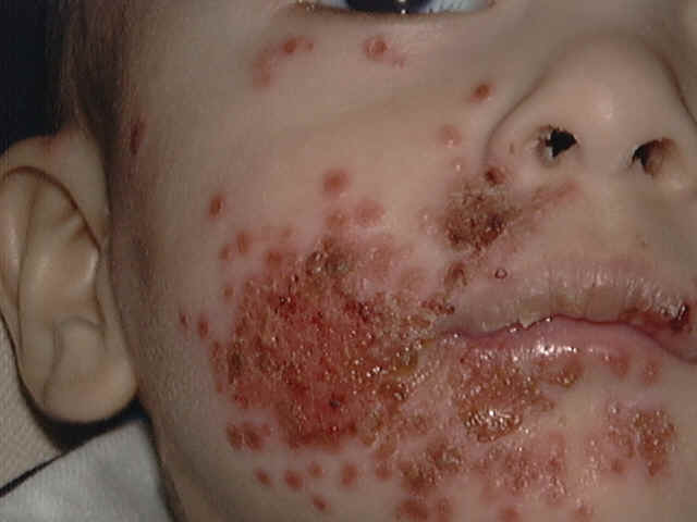
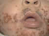 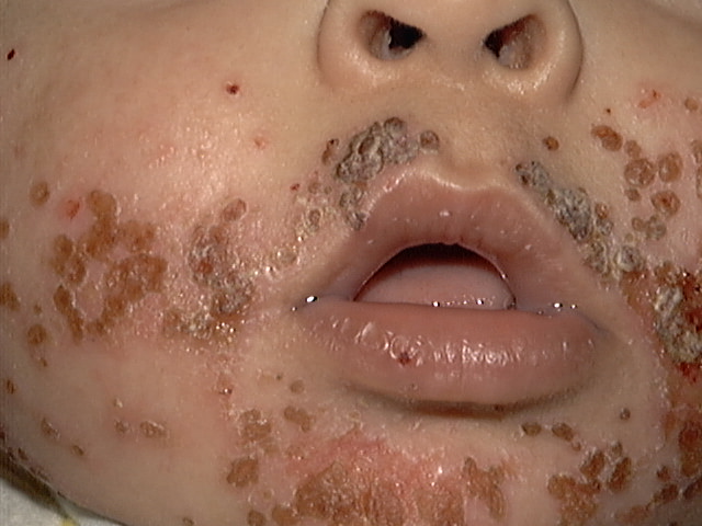
Fig.91c. Eczema herpeticum
Clinical features
Sudden appearance of umbulicated varicelliform
eruption on the sites previously involved by atopic dermatitis. The
vesicles may be hemorrhagic or complicated by secondary bacterial infection
causing more severe constitutional symptoms such as fever and lymphadenopathy.
The vesicles may
continue to appear during the course of the disease till enough
neutralizing antibodies are formed where in such cases the symptoms become
less severe , with shorter course and called the
abortive form .
N.B. Infants or
children having atopic dermatitis should not be vaccinated with small pox
vaccine due to the risk of eczema herpeticum.
The risk of
vaccination far exceeds the risk of small pox infection .
5.Fatal viraemia (generalized infection of the new born)
This is a systemic
viral infection that begins in the first week of life. This manifests with
fever, subnormal temperature, cyanosis, hepatosplenomegaly,
kidney and adrenal involvement besides the herpetic skin lesions
.
This is a severe and
even fatal herpetic lesion of the newborn caused by herpes virus type 2 due
to infection of the mother by herpes genitals. When the mother has genital
herpes during labor, there is a strong indication of delivery by cesarean
section.
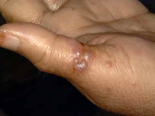
6. Herpetic
whitlow
Herpes virus may be
seeded on a wound.

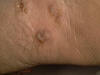
Fig.91d & e. Herpetic whitlow
Vesiculation and ulceration appears on the
infected area .
Care should be taken
not to incise the wound because the content of the vesicles are infectious
and the condition is self-limiting causing minimal symptoms.
7. Herpetic kerato-conjunctivitis
Herpes simplex may
affect the eyes causing corneal ulcers , keratitis or kerato-conjunctivitis.
The adjacent skin of the eyelids may show herpetic vesicles and ulceration.
8. Herpes progenitalis
This is a venereal
disease, which is sexually transmitted. The condition begins with burning
and tingling on the affected skin followed by the appearance of small
vesicles that tend to ulcerate . The course is
short but recurrence is common at the same site or near by in the genital area .
The common site is on
the penis and scrotum in males, vulva and vagina in females. Infected
mothers may transmit the disease to their babies during or after labor.
9.Anogenital
herpes in infants and children
Ano-genital herpes in younger age groups have number of
possible causes. Sexual abuse should be considered where thorough history
and investigations are necessary to detect the mode of infection. Direct
infection from the nursery, mother, housemaids or others may be one of the
possible causes
10. Central
nervous system herpetic infection
Rarely
herpes virus may invade the nervous system leading to encephalitis, meningeal irritation, and cranial nerve lesions with
localized neurological signs and coma. The condition may be fatal .
11.
Recurrent herpes simplex
One of the distinct
features of herpes simplex is its tendency for recurrence. It is believed
that the herpes virus becomes dormant in the tissues and flare up when
there is optimum predisposing factors.
Herpes simplex has
certain familial tendencies and infection with the virus will not lead to
lasting immunity as most other viruses .
Recurrence of the herpetic attack usually involves the same previous
location or a near by areas.
Recurrence of the
lesion may be precipitated by different factors such as common cold, fever,
strong sunlight, psychic trauma, gastrointestinal upset and menses. Almost
all humans eventually had an attack of herpes simplex during their life.
Diagnosis of
herpes simplex
Diagnosis of herpes
simplex can be established by:
·
Clinical picture.
·
Smear from the base and roof of the vesicles demonstrates
giant cells and multiple nuclei and inclusion bodies .
·
Electron microscopy demonstrates the intercellular virions characteristic of herpetic lesions
.
·
Indirect fluorescent antibody .
·
Neutralizing antibodies shows rising titer in primary herpetic
lesions.
·
IgM antibody to herpes simplex
virus
·
Tissue cultures . This is
usually expensive and rarely needed .
Treatment
It should be noted
that topical corticosteroids are contraindicated in viral disease infection
since they may cause flare up of the lesions and depress serum interferon.
Mild uncomplicated
eruptions of
herpes simplex require no treatment.
Mucocutaneous lesions may be treated simply
by 10 percent aluminium acetate or 1:8000
potassium permanganate compresses to dry the lesions .
Topical Acyclovir : every four hours is usually
enough for primary and in non-recurrent lesions. Topical Acyclovir is of
established value for herpetic keratitis.
Systemic Acyclovir : is the treatment of choice
for severe or potentially severe herpes simplex infection. Treatment should
be started as soon as possible. The usual dose is 5 mg/kg 8-hourly intravenously .
In neonatal herpes
and encephalitis: twice that dose has been used. As the drug is excreted via the
kidneys the dose must be scaled down in renal failure. Transient rises in
blood urea and creatinine may occur; slow
infusion over one hour in an adequately hydrated patient is recommended.
In the immune
compromised patient , mucocutaneous
herpes simplex respond well to intravenous Acyclovir . The infection can be
prevented by intravenous or oral Acyclovir, which should be started several
days before the anticipated immuno-suppression
and continued throughout the period of greatest risk.
The risk to the
infant from
primary herpetic vulvo-vaginitis in the mother at
the time of delivery is so great that ceasarian
section is indicated, and prophylactic Acyclovir should be considered for
the neonate .
Acyclovir orally has proven clinical value
against herpes simplex and varicella-zoster
viruses, though the latter is somewhat less sensitive to it. The usual
adult oral dose is 200 mg five times daily meanwhile,
800 mg twice daily has been used with success. The drug is given for 5 days
or more. Acyclovir is effective in eczema herpeticum
and neonatal herpes which reduces the mortality and morbidity of herpes
simplex encephalitis .
Chicken pox and
herpes zoster
dose:
* children
6 years and over: 800 mg four times daily for 5 days.
* 2-5 years : 400mg Zovirax fout times daily for five days.
* under
2 years :200mg Zovirax suspension ( teaspoonful ,
5ml ) four times daily for five days.
Herpes simplex :
* Adults and children
above 2 years : one tablet 200mg or 5 ml
suspension five times daily for 5 days.
* children
under 2 years : half the adult dose.
Recurrent attacks
of herpes simplex :
Initial eruptions of
genital herpes improve significantly by oral Acyclovir but recurrent
infections respond less well .
Frequent recurrences
can be suppressed by long-term treatment.
Prophylactic doses
vary between 200 mg and 1000 mg daily (adult doses). A typical regimen is
400 mg twice daily, gradually reduced to find the minimum effective dose
for the individual patient.
The prevention of the
predisposing factors should be considered.
Treatment of the more
severe recurrences in adults may, however, be worthwhile. In such cases it
is important to use Acyclovir tablets for longer period in smaller tapering
doses which may last for few months .
The regime that I
usually use in such cases is as follows :
Five tablets, 200 mg
daily are given for five days, then three tablets daily for another five
days, and two tablets daily for five days, one tablet daily for five days
and then one tablet twice weekly for one month and one tablet weekly for
three month. I tried this regime and gave encouraging results with severe
recurrent cases of herpes especially type 2 herpes.
Interferon: may have some effect on
recurrent herpes simplex.
Other reported
methods include topical surfactants and Cryotherapy.
Systemic Vidaribine or Phosphonoformate
for severe cases of herpes simplex infection resistant to Acyclovir.
HERPES
ZOSTER
Herpes zoster is a
vesicular viral eruption caused by the varicella
-zoster virus . A cross immunity is believed to exist
between the two diseases. Children infected by varicella
are immune to herpes zoster and vice versa.
|
Fig. 92. Herpes zoster
Herpes zoster
|
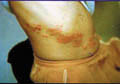
Fig. 93. Herpes
zoster
|
Clinical picture
The incubation period
is 1-2 weeks. The eruption has a rapid onset, usually unilateral and
appears along the course of nerves. The lesion is preceded by prodromal symptoms such as mild fever, pain, burning
and tingling at the site of infection.
Grouped clear vesicles on an erythematous
base appear which become purulent and rupture later on to form crusted
lesions
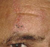 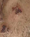
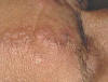
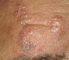
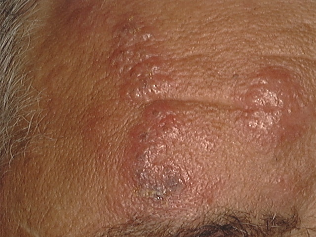 Fig.
93.d,e&F. Herpes zoster Fig.
93.d,e&F. Herpes zoster
Fig. 93b&c . post-herpetic scar
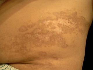 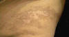 Fig. 93b. Post herpetic
scar
Fig. 93b. Post herpetic
scar
Scarring at the site
of the primary lesion usually follows healing.
In children the clinical picture may
manifest with erythema multiform-like reaction
characterized by sudden appearance of round red papules. Erythema multiforme may be
recurrent in the spring or precipitated by exposure to sunlight or after
corticosteroid treatment .
Post herpetic
neuralgia may appear after healing of the lesion accompanied with severe
pain that may be agonizing and persist is for a long time.
Herpes zoster lesions
are usually localized but generalized eruption may occur with chronic
debilitating diseases such as malignant lymphomas .
Complications
Gangrene of the zoster lesions
especially in debilitated patients.
Cellulitis and pustular
lesions due to secondary bacterial infection.
Kerato-conjunctivitis in ophthalmic lesions may
cause scarring and blindness due to progressive ophthalmic involvement.
Encephalitis and ataxia due to cerebellar disturbance is a rare complication.
Pneumonitis with cough
, dyspnea , cyanosis and scattered
calcified nodules of the lung.
Post herpetic
neuralgia is uncommon complication that is
sometimes severe and lasts for a long time causing agonizing pain.
Treatment
Potassium
permanganate
compresses 1: 8000 can dry wet oozing lesions.
Topical Acyclovir (Zovirax )cream applied
every four hours.
Disposal gloves
should be used when applying the cream by the fingers.
Oral Acyclovir - adult dose is 200mg. five
times daily for five days or 800mg twice daily. Faciclovir
250 mg. three times daily is also effective. Another antiviral preparation
is Valacyclovir 1000 mg. three times a day for
one-week. These doses are the adult dose.
Children's doses
depend on the body weight. Younger age groups can be given Acyclovir in a
dose of 5mg,/kgm body weight.
Strong sedatives sometimes are necessary to
relieve severe pain .
High doses of
vitamin B complex
may help relief of post herpetic neuralgia .
Steroid injection as ( depot
medrol 40mg. ) is believed to minimize post
herpetic neuralgia if given early . Children can be given 10-mgm depot medrol as a single injection in the early stage of the disease . Topical steroids are contra indicated in viral
skin diseases .
CONGENITAL
HERPES ZOSTER
Congenitally acquired
herpes zoster in the newborn occurs due to transplacental
infection with varicella -zoster virus . This is a serious problem that produces
congenital abnormalities . Congenital varicella zoster may be acquired by transplacental
varicella-zoster virus infection
. The manifestations that appear after birth are cutaneous
scars , limb and eye abnormalities . The
manifestations are serious if infection occurs in late pregnancy
.
Treatment
Acyclovir is given in
a dose of 1000 mg./day orally for five days .
Topical Acyclovir is applied to the skin or the ophthalmic lesions repeatedly .
CHICKEN
POX
Chicken pox is a
highly infectious viral diseasecaused by the varicella - zoster virus. Children are the most common
age group infected. A rash that has a central distribution characterizes
the disease, which occurs in widespread infection and occurs in epidemics
especially in schools and crowded communities. Usually there is lasting
immunity for varicella and herpes zoster ,however zoster may occur sometimes after a varicella infection.
Clinical Picture
The incubation period
is from 1-2 weeks . Transmission is usually by
droplet infection, direct contact with the lesion or from recently contaminated
fomites.
Systemic
manifestations:
The disease presents
with a mild attack of sore throat , fever ,
headache that lasts for 2-3 days . This is followed by the appearance of
the characteristic rash on the trunk and mucous membranes, which may become
generalized .
Skin
manifestations:
Skin rash manifests
with erythematous macules,
vesicles and pustules which rupture leaving crusted lesions. The rash is pleomophic where different stages of the rash , macular , vesicular and pustular
lesions of different sizes are present at the same time .
The crust may
separate after one week leaving in severe cases scars and hyperpigmentation especially in the dark skinned patients .
The lesion has a
characteristic central distribution ; on the trunk
more than on the extremities .
Itching is usually
mild but may be severe in some cases .
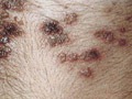
Complications
The clinical picture
is usually mild but may become severe mainly in adults ,
involving the skin and mucous membranes associated with fever and severe
constitutional symptoms .
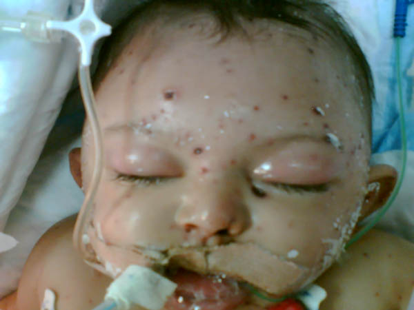
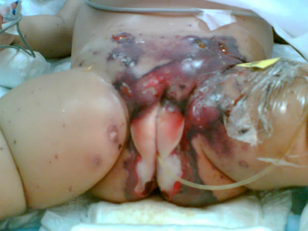 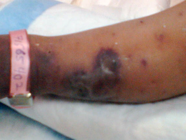
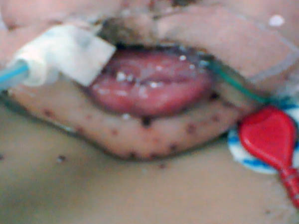 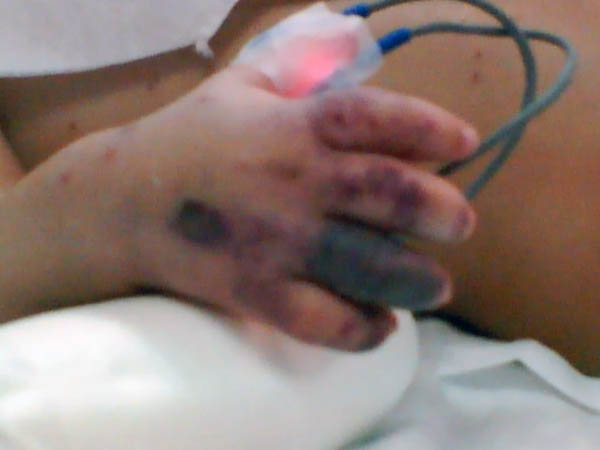 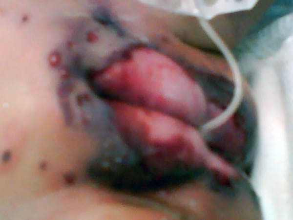
Fig.
93 c,d,e,f,g,h. Complicated chicken pox (Caurtesy of Dr. W.Khalaf - R.K.H
-K.S.A)
Encephalitis, meningeo-encepalitis and pneumonia are uncommon
complications of the disease.
|
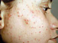
Fig. 94.
Chicken pox
|
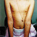
Fig. 96-.
Chicken pox
|
|
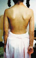
Fig. 95.
Chicken pox
|
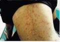
Fig. 97-.
Chicken pox
|
Diagnosis
The diagnosis of
chicken pox depends on different data mainly:
·
Characteristic clinical picture.
·
The centripetal distribution of the skin lesions.
·
Pleomophic different stages of the
eruption.
·
Laboratory investigations.
Differential
Diagnosis
Small pox
Eczema herpeticum
Pustular impetigo
Drug eruption
Treatment
Children should not
go to school until complete healing of the lesions.
Mild cases can be
treated with mild soothing agents.
Calamine lotion,
which is commonly used to dry the lesions and minimize itching, but
excessive use can cause more dryness and irritation.
Weeping wet lesions
are better treated by Potassium permanganate compresses 1:9000 applied
twice daily .
Non-steroid
anti-inflammatory topical cream such as Pufexamac
(Droxaryl Cream) may be applied. This medication
is safe and we found out that it is very effective in rapidly clearing the
skin lesions and relieving itching.
Antihistamine orally
may be needed for relieving of itching.
Severe cases
especially in adults, may need isolation and
hospitalization till the severe eruption, constitutional symptoms or
complications are controlled.
NEONATAL
VARICELLA
The risk of fetal
infection with varicella occurs when a pregnant
woman develops chicken pox 3 weeks before delivery .
Clinical
Manifestations
The manifestations,
prognosis and the severity of the disease depend on the onset of infection
in the mother and infant.
1.
Pre-natal infection
a.
Mild neonatal infection - the infection is usually
mild if the onset of the disease in the mother is in the first week before
delivery and within the first 4 days in the neonate.
b.
Severe and disseminated neonatal infection - this may occur if the
mother is infected within 4 days before delivery and the neonate is
infected in the first 5-10 days after delivery.
The infection is likely to be disseminated and severe, with
involvement of the lungs, liver and the brain. In these cases there is
usually a high mortality rate.
c.
Congenital varicella
syndrome
- Intra-uterine varicella infection in the first
trimester may result in a characteristic combination of defects in the
neonate known as the "congenital varicella
syndrome." Limb hypoplasia and zosteriform cutaneous
scarring are common manifestations.
2.
Post-natal infection
Post-natal
acquired varicella that may be more severe in the
following cases:
If
exposure occurs in the first few days of life, particularly in
infants whose mothers had not previously been infected and therefore did
not provide passive immunity.
Premature
infants in whom very little passive immunity have been transferre
General
Manifestations
Headache, nausea,
vomiting, fever, nucheal rigidity, and rarely
seizures.
Central nervous
system involvement occurs in less than 1% of cases of varicella.
Cerebellar ataxia and Parkinson-like
features are the most common presenting neurological signs
.
Skin
manifestations
The same as the
ordinary varicella in different age groups. The
lesions may be severe and the rash is more widespread and extensive.
Diagnosis
Distinction between
neonatal varicella and herpes simplex virus
infection can be reliably confirmed only by viral culture.
A history of genital
herpes in the mother .
The presence of kerato-conjunctivitis .
Typical herpetic oral
lesions.
Treatment
The condition may be
serious. It may endanger the infant‘s life.
Hospitalization and
proper nursing is of prime importance .
Zoster-immune
globulin or gamma globulin should be given to neonates born to any mother
who develops chicken pox during the last 4 days of pregnancy.
THE INFECTIOUS EXANTHEMATA
Infectious
exanthemata include viral diseases that are characterized by exanthematous skin lesions, fever and systemic
manifestations. The problem may pass without disturbing the child health or
may be severe endangering his life .
Exanthemas include
mainly measles, rubella, roseola and infectious mononucleosis .
MEASLES
Measles is an endemic
viral disease . The most majority of the population have had the disease. Measles is infectious
during the prodromal stage, which is followed by
the skin rash.
Clinical
Feature
Prodromal symptoms:
The incubation period
is from 2-3 weeks . The lesions begin with a prodroma of fever up to 39*C, headache, malaise, sore
throat, coryza and conjunctivitis. The early
manifestations are not characteristic at the beginning and may be confused
with
influenza.
The symptoms subside
as the rash develops.
Skin
manifestations
Skin rash begins few
days after the prodroma where the symptoms may be
more severe and the child may become seriously sick .
Rash appears first on
the forehead and is characteristically more dense behind the ears which
later involves the face, trunk and extremities. The temperature then
returns to normal following spread of the rash .
Mucous membrane
involvement
One of the main
characteristic of measles is the Koplik‘s spots
that appear as small bluish white lesions on an erythematous
base around the orifices of the parotid duct and occasionally on the lower
lip.
Conjunctivitis and
photophobia are common manifestation of measles .
Lymphadenopathy may be present but usually it
is not a marked feature.
Severe cases may show
serious complications such as encephalitis .
Treatment
The different lines
of treatment are general and these include the following: Palliative
treatment
Bed rest and
protection from exposure to strong sunlight .
Symptomatic treatment
mainly for cough .
Antibiotics
: If there
is secondary bacterial infection .
Corticosteroids may
be needed in cases complicated by encephalitis .
Light food and fluids
are given especially during the prodromal stage .
Hospitalization in
severe and complicated cases of measles where concentrated plasma globulin
may be given.
GERMAN MEASLES
( Rubella )
German measles is a
very mild exanthematous disease. If a pregnant
women is infected during the first trimester, serious fetal malformation
may develop such as deafness, cardiac malformation, cataract, microcephaly and dental malformation
.
Usually miscarriage
is medically indicated if these abnormalities are confirmed
.
Clinical Features
Prodromal Manifestations
Prodromal symptoms may be very mild and
usually pass without notice .
Enlargement and
tenderness of lymph glands begin 5-7 days before the rash appears. The
enlargement is generalized but characteristically involves the suboccipital, postauricular
and cervical glands but this is not pathogonomonic
for rubella. The tenderness of the glands subsides after a day or two but
palpable enlargement may continue for several weeks.
Skin
Manifestations
Skin eruption present with fine round pink macules that appear on the face, head and trunk which
persist for 2-3 days and then disappear .
Mucous Membrane
Manifestations
Dull-red macules or petechiae may be
detected on the soft palate, but Koplik‘s spots
are not detected .
Complications
Arthritis is not
uncommon, involving small joints of the hands feet or knees, elbows and
shoulders.
Purpura, thrombocytopenic or non-thrombocytopenic .
Encephalitis is very
rare.
Treatment
Usually the condition
resolves within few days and no treatment is required.
Vaccination with the
rubella virus vaccine for females in the child-bearing age is necessary,
taking much care that the female is not pregnant.
CONGENITAL
RUBELLA
Mothers who have had
rubella during the first trimester of pregnancy may give birth to infants with
a syndrome triad consisting of :
·
Congenital cataract
·
Cardiac defects
·
Deafness
Skin
Manifestations
Cutaneous lesions are among the most
prominent clinical features of congenital rubella.
The typical lesions
are present at birth or appear during the first 48 hours. The skin rash is
discrete, rounded, red or purple infiltrated macules,
3-8 mm in diameter. The lesions are mainly on the face, scalp, the back of the neck and on the trunk.
Occasionally the
lesions are slightly raised. They tend to fade over a period of weeks.
These lesions have often been described as ‘purpuric‘ and have generally been attributed to
thrombocytopenia, which is another common feature of congenital rubella .
Systemic
Manifestations
Disseminated
infection of rubella causes intra-uterine growth retardation, microcephaly, micro-ophthalmic and a wide variety of
other serious manifestations.
Treatment
No specific
treatment.
Symptomatic treatment
for the accompanying manifestations .
RUBELLA
IN PREGNANCY
This is a serious problem,
if infection occurs during the first trimester of gestation. Prenatal
damage with risk of fetal abnormalities occur in
most cases. Intra-uterine infection leads to malformation of the fetus.
Heart and eye damage
is most frequent in embryos infected under 6 weeks.
Deafness and mental
deficiency occurs in embryos of all ages up to about 16 weeks.
Mental retardation
and microcephaly may not be apparent until a year
or more after prenatal infection .
ROSEOLA
Roseola is an exanthematous
viral disease affecting babies and young children.
Clinical features
Prodromal symptoms
The disease usually
presents with variant and grave manifestations such as fever, convulsions
and lymphadenopathy. The child may be seriously
ill. On the fourth or fifth day the fever suddenly drops and the child
general condition is improved where he becomes active and has an increased
appetite.
Skin
manifestations
Rose-colored discrete
macules appear after the drop of fever . The common sites for the rash is the trunk, neck , buttocks , extremities and to less extent on the
face . The rash may persist for few days where it vanishes gradually.
Mucous membranes are
not involved .
ROSEOLA
INFANTUM
(Exanthema subitum)
Roseola infantum
is a common exanthematous disease of babies and
young children almost restricted to the first 3 years of life. The disease
is believed to be due to Coxsackie virus .
The incubation period
is about 10 days and is characterized by sudden onset of high fever that
usually subsides with the onset of the skin eruption.
Clinical Features
Prodromal manifestations -
High fever,
convulsions.
Sudden drop of fever.
Within four days the
child who has been severely ill sits and resumes his activities.
Skin
manifestations
Skin eruption appears
when fever begins to subside .
Periorbital edema and hematuria
are usually the early manifestations.
The skin rash is morbilliform erythema
consisting of discrete rose-pink maculopapules
that appears first on the trunk with mild involvement of the face . The skin lesions may become extensive where it
may spread to the neck , arms, and legs. After 1
or 2 days the rash fades, leaving neither scaling nor pigmentation.
Mucous membranes are spared from any lesion .
Cervical and
occipital lymph nodes are usually enlarged.
Systemic
manifestations
as febrile convulsions are not uncommon but encephalitis is rare. The
disease is uniformly benign.
Differential
Diagnosis
Roseola may simulate different skin
conditions such as measles and drug eruption . The
disease can be differentiated from measles by the absence of prodromal respiratory symptoms ,
distribution of the eruption and the absence of Koplik‘s
spots .
SMALL POX
(Variola Major)
Small pox is a highly
infectious viral disease that has a high mortality rate and occurs in
epidemics. Different strains of small poxvirus have different virulence and
variable clinical manifestations.
Clinical Picture
The clinical picture
of small pox may vary greatly . The incubation
period is about 12 days .
Skin manifestations
The cutaneous lesions may show hemorrhagic pustules or erythema multiforme like
eruption.
Sudden onset of fever
and malaise with an exanthematous papular, vesicular, pustular
and crusted lesions involving characteristically the face, extremities, palms
and soles.
Secondary bacterial
infection may cause, secondary pyogenic infection
of the skin .
Bronchopneumonia
Corneal ulcer .
Encephalitis
.
Diagnosis
·
Microscopic examination of skin
scrapings of fresh vesicles show elementary bodies.
·
Electron microscopy.
·
Culture of the scrapings or the contents of the vesicles .
·
Complement fixation test.
Treatment
No specific treatment
against small pox has been developed .
Cases are isolated in
a designated small pox hospital .
Complicated cases by
secondary infection can get benefit from antibiotics. Corticosteroids may
have a value in encephalitis.
Symptomatic
palliative treatment.
Preventive measures
by small pox vaccination .
However, routine
vaccination of children is no longer necessary in areas where the disease
is eradicated, while in certain areas infected with small pox vaccination
is necessary.
VACCINIA
The vaccina virus is an attenuated Cow box virus that has
been propagated in laboratories for small pox vaccination. Skin lesions due
to vaccinia virus result from complications of
vaccination against small pox .
The manifestations
appear in different clinical pictures :
Immediate response
or immune response : A papule appears immediately
after vaccination that involutes after the third day of vaccination.
Primary response : In the third day of vaccination a papule appears that
becomes a vesicle on the ninth day then a pustule . The condition is
accompanied by regional lymphadenopathy.
Accelerated response : A papule appears on the fifth day and then a small
vesicle is formed which involutes on the ninth day.
Generalized vaccina : generalized papulo-vesicular eruption may follow 10 days after
vaccination. The eruption then changes to pustular
type, which may involute within three weeks . Successive crops may follow .
Retinitis and ocular paralysis may complicate the condition
.
Eczema vaccinatum : This is the same as eczema herpeticum , which occurs with herpes simplex due to
inoculation of the vaccine at sites of atopic dermatitis.
Multiple vaccination : Multiple skin sites may be involved due to contact of
the skin from vaccinated site of the same individual or contact with other
vaccinated .
Roseola vaccinia : Infants and young children
are mostly affected. The skin rash is morbilliform
eruption. The vaccinated area becomes crusted and surrounded by an erythematous halo, which involutes within few days.
Vaccina necrosum : This occurs in infants under
six months of age that are unable to produce antibodies in response to
vaccination . Necrotic metastatic lesions occur throughout the body. The
condition is usually fatal .
Roseola vaccinia : Symmetrical discrete papular , macular and morbilliform
eruption appears two weeks after primary vaccination with small pox
vaccine. The site of vaccination becomes crusted and surrounded by an erythematous halo .
HAND - FOOT -
MOUTH DISEASE
This is a viral
disease caused by Coxsackie virus 16 affecting mainly children.
Clinical Features
Prodromal symptoms: fever precedes the appearance
of the rash.
Skin manifestations: a striking
features of this disease is the appearance of oval, linear or
crescent maculopapular eruption that shortly
becomes vesicular on the hands, feet and in the mouth. The eruption, which
appears on the hands and feet, is usually parallel to the skin lines.
Spontaneous recovery
is usually within two weeks and treatment may be not needed
.
FOOT - AND - MOUTH DISEASE
This is a highly
contagious viral disease that has an incubation period from 2-10 days. It
is transmitted to man directly from infected animals such as cattle, goats
or from consuming their infected milk . The
disease may have serious complications and sometimes may be fatal
especially in children .
Clinical Features
Prodromal symptoms are mild. This
includes fever, malaise, burning and dryness of the mouth with excessive salivation .
Skin eruption appears
with the decline of fever and disappearance of the prodromal
symptoms.
Skin and mucous
membrane manifestations : Swelling , itching and burning
sensation of the fingers may be the earliest skin manifestations . Vesicles
then appear on the mouth , oropharynx
, palms ,soles , fingers and toes.
Treatment
Symptomatic
treatment.
HIV
DISEASE IN CHILDREN
HIV disease in
childhood in many respects has the same manifestations such as that of the
adult type.
Clinical Features
The most common
manifestations of HIV in infected infants and children are:
·
Failure to thrive.
·
Encephalopathy with developmental delay.
·
Chronic parotid swelling .
·
Hepatosplenomegaly.
·
Lymphadenopathy .
·
Chronic diarrhea.
·
Bacterial infections: septicemia, pneumonia, otitis media and cutaneous
infection mostly due to Staph. aureus
causing impetigo, abscesses and cellulitis.
·
Pneumonitis: which may be due to
infection with Epstein-Bar virus , is common and
characteristic.
·
Malignancy :Kaposi‘s sarcoma occurs in
only about 5%.
Laboratory
diagnosis
1.
B-cell dysfunction: is an important early finding .
2.
Circulating HIV antigen :is
likely to be obscured by maternal antibody.
3.
T-cell defects: characteristic of adult HIV disease
are usually a late feature in young children
4.
HIV antibodies - the presence of HIV antibody IgG in infancy may simply represent passively acquired
maternal antibody, and cannot be used as an indication of HIV infection.
Prognosis
Some infected
children develop AIDS within their first year. In the majority of cases,
the infection progresses more slowly .
Treatment of HIV
infection in childhood
Treatment of complications .
Treatment of
secondary bacterial infections mainly pneumocystis
pneumonia.
Antiviral drug
therapy at present cannot cure the infection.
The principal agent
is zidovudine (Azidothymidine,
AZT, Retrovir).In spite there is imptovement of some cases, but these must be weighed
against the drug toxicity including anemia, granulocytopenia,
myositis, headache, confusion, insomnia, nausea,
fever, rash and nail pigmentation.
Zidovudine is usually given orally for
prolonged periods. Occasional cases of resistance have been reported. Serum
and CSF HIV antigen levels can be greatly reduced and may become
undetectable.
Long-term intravenous
infusion of Zidovudine may lead to improvement of
childhood neuro-developmental abnormalities
.
There is no available
vaccine or specific treatment now for HIV,
meanwhile efforts are tried in the different international centers to find
a curative treatment.
Prevention
Prevention of
transmission of HIV is therefore of paramount importance. The guiding
principle is to avoid contact between infected secretions and mucosal
surfaces or broken skin.
HUMAN IMMUNODEFICIENCY
DISEASE
IN BREAST-FED INFANTS
There are reports of
occasional cases of apparent transmission in breast milk. In one case a
baby probably acquired HIV from an infected wet nurse. The risk is believed
to be low compared with that of pre- or intrapartum
transmission in such cases .
WARTS
(Verruca Vulgaris)
General
Considerations
Warts are extremely
common viral infection of the skin of children and young adults. They have
different morphological characters concerning the shape, size and sites
involved. They may be single or multiple, sessile with rough hard surface
or flat as those on the face. Warts may be dry such as warts of the skin
surface or wet as those on the anogenital areas .
Etiology
The human wart virus
causes warts that belong to the Papovavirus,
which contains DNA. Warts are contagious viral infection. Auto-infection is
common especially in children who have warts of the fingers and used to put
the finger in mouth or bite an area infected with warts. This may cause
auto-infection to lips.
Direct contact of the
infected area to the traumatized skin surface may cause infection.
Morphological
types
The common warts
are raised, dry lesions with rough gray surface. The most common sites involved
are extremities particularly the fingers.
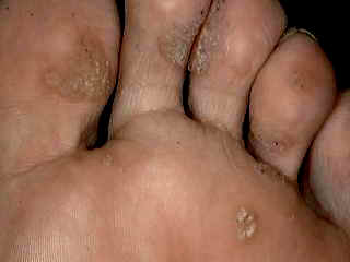
Plantar warts are inverted warts due to
continuous pressure during walking. Such type is
not raised as other warts. Warts may be single or multiple and may
be painful if they are located on the pressure sites.
|
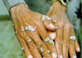
Fig. 98-. Verruca Vulgaris
|
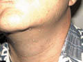
Fig. 99-. Filiform Warts
|
|
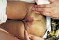
Fig. 100. Anal & Perianal Warts
|
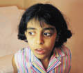
Fig. 101. Warts of the
eyelids
|
|
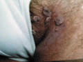
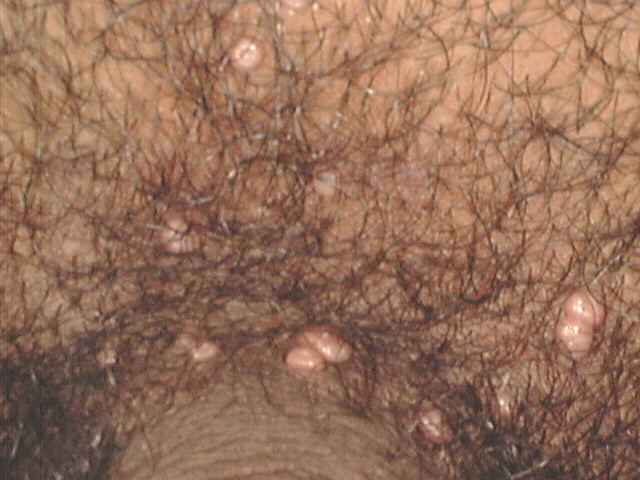
Fig. 102. Moist crural warts
|
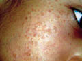
Fig. 103. Flat Warts of
the face
|
|
[AD-SIZE]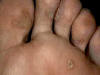
|
Fig. 104. Plantar Warts
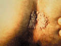
Fig. 105. Fungating warts |
|
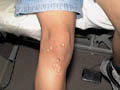
Fig. 106. Warts of the knee |
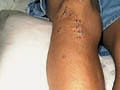
Fig. 107. Warts treated by Co2 Laser |
|
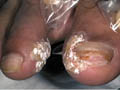
Fig. 108. Periungual Warts treated by
salicylic & Lactic acid |
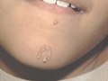
Fig. 109. Warts (Koebner's Phenomond) |
Flat
warts : Located mainly
on the face and sometimes are not easily differentiated from freckles.
Filiform warts : Located
mainly on the neck and face which are differentiated from cutaneous horns
and skin tags by being harder than skin tags .
Moist ano-genital warts :
Common on the glans penis in males and vulva in females besides the anal
area or the skin of the anogenital area.
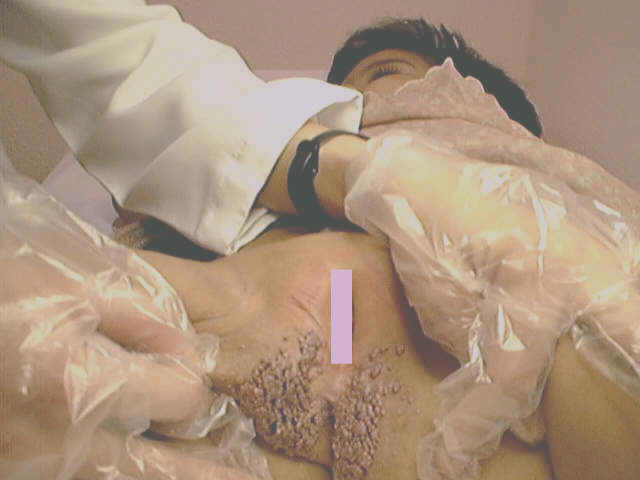
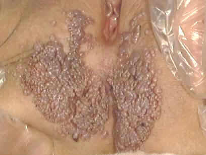
Fig.100a&b.
Ano-genital warts (Condyloma accuminata),(before treatment)
(Recurrence after
expensive and unsuccessful surgical excision besides different
topical medications for the last three months in other medical
centers. The father claims that cost was more than 3500 $ !!
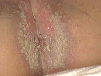
Fig.100c.
Photo of the same infant treated in our medical center after three
applications of 20% topical Podophyllin in Benzoin co , one application every two
days and washed after four hours . ( The cost of that treatment was only THREE DOLLARS !!!!!
).
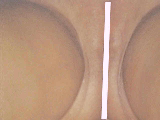
Fig.100.d&e.The same child after 10 days(she
was given mupericin cream (Bactroban cream) applied once
daily .
N.B: Human papillomavirus (HPV)
is the causative of genital warts.
Treatment
Different treatment
regimes have been used for treatment of warts since a long time. Some of
these regimes are traditional used by non-medical personnel such as
religious persons reading from the holy book to the infected individuals
or to occlude warts by caustics for sometimes or by suggestion .
Treatment of warts depend
on different factors .
There is a different
alternative methods for treatment .
-
Common warts can be
treated by liquid nitrogen, Electrodessication using local infiltrating
anesthesia . It is important to dissecate the base of the wart but not
to go deeper where the warts are intraepidermal in order not to leave
much scarring after electrodessication.
-
Moist anogenital
warts can be treated effectively by 15-25 percent Podophyllin in
collodion or in tincture Benzoin to be applied cautiously to the
affected areas and washed after 8 hours . Application may repeated after
three days or one week, where after the warts slough .
-
Peri ungual warts
are difficult to treat. Electrodessication may be
used but recurrence is
common.
Different lines of
treatment are known.The cheapest and most reliable is Podophyllin
resin in different concentrations ,(10-25%) in Tinc. Benzoin co. or
collodion ( applied every
other day or twice weekly ) and washed after 4-6 hours.
Other topical medications are
Immiquimod (Aldara
cream and Podofiliox ( Condylox gel 0.5
)
The recent and
effective treatment for warts is by CO2 Laser.
We use CO2 laser for
treatment of most reluctant and extensive warts such as peri ungual ,
moist warts and plantar warts using topical (Emla cream as an anesthetic
preparation ).
-
Plantar warts - we
use a formula containing the following : salicylic acid 20 per cent, lactic acid 20
percent in flexible collodion. This preparation is very effective in
treatment of plantar warts and in common warts especially in children
who refuse other methods, which may need local infiltrating anesthesia.
This preparation should
be used with great care to the wart area and to be used by the mother
for her child because it may cause severe chemical burn if it comes in
contact with sensitive parts of the skin.
When this preparation is applied to
plantar warts, it causes gradual exfoliation of the skin, so daily before
applying the medication , the area is scrapped or shaved to remove the
dead skin . Black or gray spots can be seen these represent the thrombosed
vessels. Application is repeated on these areas, where usually after one
week to ten days the patient begins to feel deep-seated pain on applying
the medication. This indicates that the medications reached the tip of the
inverted wart. Another two applications may be enough to reach a curative
stage for the plantar warts .
-
Facial warts can be
treated by liquid nitrogen or by salicylic 5 % , lactic acid 3 % in
collodion preparation with different strengths according to the age of
the patient and type of skin. Young children may need less concentrated
preparations as 3% salicylic, 3 % lactic acid in collodion .
EPIDERMODYSPLASIA
VERRUCIFORMIS (EV)
Epidermolysis dysplasia
is a viral infection caused by the human Papovirus
Clinical Features
-
Skin lesions:
Symmetrical lesions characteristically appear on the extremities, dorsum
of the hands and feet, neck and face. The lesions are dry, rough, flat
well-defined papules simulating verruca vulgaris.
-
Mucous membrane
lesions may appear on the anogenital area and on the lips.
-
Nail dystrophy
-
Hyperkeratoses of the
palms and soles may accompany some cases.
-
The disease may be
complicated by malignancy such as epidermal carcinoma in an early age.
Treatment
Traditional treatment as
for warts is usually not possible to eradicate all lesions since theses
are numerous .
CO2 Laser can be used successfully to
ablate the lesions using local anesthesia (Emla cream) under occlusion
method .
SAND FLY FEVER
Sand fly fever is a viral
disease transmitted by the female sand fly (Phlebotomus papatassii) found
in the Mediterranean area. The disease is characterized by fever,
headache, shaking chills, back pain, muscle ache and fatigue .
Clinical Features
Skin manifestations
Small pruritic nodule
appears after few days at the site of the sand fly bite. Scarlitiniform
eruption appears on the face and neck.
Constitutional symptoms
Fever, headache, malaise,
nausea and abdominal pain .
Systemic manifestations
Eye involvement:
conjunctival injection
Stiffness of the neck .
The disease has a chronic
course where recovery may occur , but relapsing attacks of fever may
continue for long time .
Treatment
Non-specific treatment of
symptoms .
DENGUE
(Break bone fever)
Dengue is a viral
infection caused by dengue virus. The disease is transmitted by Aedes
aegypti mosquito that is the vector of the organisms . The disease is
endemic in the Mediterranean areas , Africa ,Hawaiian and Caribbean
islands .
Clinical Features
The disease is
characterized by fever, headache, shaking chills, back pain, muscle ache
and fatigue.
In childhood the usual
infection is asymptomatic, or there may be mild fever, sometimes
accompanied by a rash.
In adults a biphasic
fever with headache, severe backache and a rash is more characteristic.
Skin Manifestations
Maculopapular or
scarlatiniform rash appears on the third to fourth day of the fever. It
starts on the chest and trunk and spreads to the face, arms and legs. The
rash fades as the fever subsides but can be followed by petechiae on the
arms and legs. In dark-skinned people the rash is frequently not visible.
Systemic Manifestations
Hemorrhagic
complications: petechiae, which can be demonstrated by a positive
tourniquet test (Hess test ). This typically occurs in children who have
had a previous dengue infection of a different serotype.
Body temperature falls
within one week and shock ensues, where at this stage the patient may die.
Pleural effusion and
ascitis
Diagnosis
Different criteria may be
of help in the diagnosis of the disease. These include the following:
-
Typical clinical
picture such as fever chills and aching pain .
-
Skin eruption is of
the morbilliform type or exanthematous involving the face, neck and
chest.
-
Confirmation is
obtained by culture of blood in the acute phase.
-
Serological studies
on acute and convalescent sera.
Treatment
The disease has favorable
prognosis and treatment is only symptomatic.
INFECTIOUS MONONUCLEOSIS
Infectious mononucleosis
is considered as viral infection perhaps due to EB virus.
Clinical Features
Systemic manifestations
Prodromal symptoms :
fever , headache and malaise .
General manifestations:
splenomegaly, lymphadenopathy mainly the cervical and to a less extent the
axillary and inguinal lymph nodes .
Skin manifestations
Skin rash appears in one
third of cases. The skin lesion is an erythematous macular eruption on the
upper extremities and trunk . Rarely scarlatiniform, urticarial or
morbilliform eruption may be seen with edema of eyelids.
Mucous membrane of the
buccal cavity may show distinctive multiple pinhead-sized petechiae .
Diagnosis
-
Paul-Bunnel test is
positive with a titers of 1:112 or higher .
-
Blood picture -
Lymphocytosis with abnormal large lymphocytes and leucocytosis.
-
Liver function tests
may show elevated SGOT and SGPT.
MOLLASCUM CONTAGIOSUM
Mollascum contagiosum is
a common viral disease in school age and in adults. Lesions involve
usually the skin and to a lesser extent mucous membrane of the mouth and
tongue.
Modes
of Infection
Transmission of the viral
infection occurs from:
-
Swimming pools
-
Infected fomites.
- Autoinoculation.
Clinical Feature
The common sites involved
are the face, hands, trunk and genitalia. The eruption may be single,
multiple, localized or generalized and has a chronic course .The
incubation period is 2-4 weeks.
|
Fig. 110. Mollascum contagiosum |
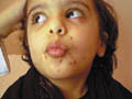
Fig. 111. Mollascum contagiosum |
The primary lesion of
mollascum contagiosum can be easily diagnosed. The papules appear as flesh
colored, solid then become pearly white, soft, rounded, dome shaped
papules with central umbulication and contain caseous plug. The papules
may suppurate due to secondary bacterial infection.
Different morphological
patterns may follow the course of mollascum contagiosa:
Giant form : The papule
may reach a huge size; more than 10 cm. which may suppurate and is
confused in the early stage with verruca vulgaris, kerato acanthoma and
basal cell carcinoma.

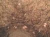
Fig. 111b. Giant Mollascum Contagiosum
Mollascum contagiosum
cornuatum : The lesions are horny, small papules.
Generalized form :
Extensive wide spread lesion involving face , trunk, extremities and
genitalia . The mouth as well as the tongue may also become involved.
Diagnosis
Mollascum contagiosum can
be easily diagnosed by the distinctive umbulicated pearly papules.
Histopathology shows
acanthoma with downward proliferation of the ret ridges.
Basophilic Mollascum
inclusion bodies are detected in the cytoplasm of the ret mucosum.
Treatment
Curettage - is the
easiest and most reliable. The lesions are sprayed with Ethyl chloride
until it becomes white freezing and then scrapped with curette.
Electrodesiccation -
certain lesions, such as the eyelids especially in children can be removed
by electro-desiccation using infiltrating local Xylocaine anesthetic.
When infiltrating
anesthesia is not possible due to irritable child , topical (Emla) cream
can be used .The cream is rubbed to the area and thick layer of the cream
is applied and occluded by cellophane cover for about 40 minuets then the
lesions can be easily curetted .
Topical tincture iodine
and cantharidin is used by others to treat mollascum contagiosum .
We use a paint containing
10% Salicylic acid and 10% Lactic acid in flexible collodion .This
preparation is effective and can be used for treatment of infants and
young children who can not afford other lines of treatment.
KAWASAKI SYNDROME
(Mucocutaneous lymph node
syndrome (MLNS)
Kawasaki and co-workers
in Japan introduced this syndrome which affects mainly young children is
of unknown etiology, in 1967. It is typically sporadic and occurs
throughout the world but is most common in Japan. There is no evidence of
person-to-person spread.
Etiology
Many infectious agents
have been suspected as the cause, including streptococci, staphylococci,
rickettsia and viruses, but in most the etiology remains unknown.
Cytokines released from monocytes affect vascular endothelial cells of
which make them susceptible to damage by circulating cytotoxic antibodies
.
Clinical Features
The manifestations of the
syndrome are mainly diffuse vasculitis .
Systemic manifestations
The onset is acute with a
remittent fever that lasts more than 5 days. The patients look toxic.
Mucous membrane
manifestations
Mucous membranes of the
conjunctiva, mouth and tongue may be involved. This may lead to
conjunctival injection, dry red lips and mouth. "Strawberry
tongue" similar to that seen in streptococcal disease . These
features continue while the fever lasts.
Skin manifestations
Children may show
perineal eruption on the perineal and crural areas covered by the diapers
.
A generalized polymorphic
rash develops. This appears as urticarial, scarlatiniform, and
morbilliform, macular, papular within 1-5 days of the onset of the fever.
It is mainly on the trunk and proximal extremities and lasts for up to a
week. Palms and soles become erythematous red, indurated and later show
desquamation.
A sub acute phase of the
illness follows the end of the fever. This is characterized by
desquamation of the skin of the fingers and toes which begins at the
skin-nail junctions
Cervical lymphadenitis is
present in 50-80% of patients..
Arthralgia and arthritis
may present at this time, but in less than half of the patients, typically
that of knees, hips and elbows.
Cardiovascular
manifestations :
These are the most
serious complications and sometimes fatal. The manifestations are
myocarditis, aneurysm, stenosis or obstruction of the coronary arteries .
In most cases, recovery
takes place slowly and is usually complete within 10 weeks.
Treatment
-
Gamma globulin: a
daily infusion of 400 mg/kg is recommended .
-
Symptomatic
treatment: Acetylsalicylic acid (Aspirin ) for the fever and arthritis.
-
Systemic steroids :
may be life saving in systemic in cardiac manifestations.
PITYRIASIS ROSEA
Pityriasis rosea is a
papulo-squamous self-limiting disease of unknown etiology. Recently a
viral infection is suspected to be the cause. Most of cases are seen in
autumn. The commonest affected are adults but children and rarely infants
may have the disease .
In our observations we
found a large number of patients who give a history of appearance of the
eruption after using new clothes and bed coverings few weeks before the
onset of skin manifestations .
Clinical Features
General manifestations:
-
Mild symptoms as
headache and slight malaise.
-
Slight fever, malaise
.
-
Enlargement of lymph
glands, generalized or confined to the cervical glands, may be present.
Skin manifestations
Herald
patch
Usually a single lesion
may precede the skin eruption . Herald patch appears mainly on the chest,
trunk or extremities in the form of large, rounded or oval , bright red
patch with well defined edge and covered by fine scales.
|
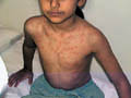
Fig. 112. Pityriasis rosea (Typical distribution
along the rib lines) |
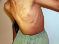
Fig. 113. Pityriasis rosea |
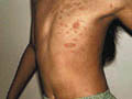
Fig. 114. Pityriasis rosea (Herald patch) |
The scales are
characteristically more on the periphery , attached at the edges and loose
towards the center. Within one week a skin eruption appears mainly on the
trunk, back and the chest characteristically arranged along the lines of
the ribs. The lesions are discrete, oval, dull pink color macules and
patches covered by dry scales . The center of the lesions may appear
hypopigmented . Pruritus is usually absent unless there is irritation of
the lesions by excessive bathing , sweating or other local irritants . The
skin lesions commonly fade after 3-6 weeks, but some clear in 1 or 2 weeks
and a few persist or as long as 2-6 months leaving no trace .
Different Clinical
Varieties
Different morphological
forms of pityriasis rosea may be seen in children and adults. In
children the lesions may be papular or urticarial in the early stages.
In adults typical medallions studded with purpuric points, and
acutely purpuric lesions are manifestations of rare types of pityriasis
rosea.
The herald patch
is absent or undetected.
Papulovesicular,
vesicular and even pustular forms may occur, and erythema
multiforme-like lesions .
Differential Diagnosis:
Tinea corporis: The
lesions show central clearing with an active edge. Skin scrapping will
show the causative dermatophyte.
Psoriasis. The lesions
show silvery scaly patches. The scales covers the whole patch.
Treatment
Pityriasis rosea is a
self-limiting disease . Complete healing without treatment of the lesions
usually takes from 2 weeks to 6 months .
No treatment is usually
needed .
Rarely the symptoms may
need antihistamine and mild topical steroid for few days to relieve
itching .
When there is distressing
symptoms especially with extensive lesions, a topical steroid, usually of
moderate strength or ultraviolet (UVB) may be used.
Usually we do not give
treatment for pityriasis rosea and the best is to avoid skin irritation
and minimize bathing , chaffing and excessive sweating. In extensive
lesions accompanied by pruritus, we give pufexamac cream for one week.
RESPIRATORY SYNCYTIAL
VIRUS
This is a viral disease
that may spread in epidemics in winter. The disease is caused by
pleomophic small virus affecting mainly children in the pre-school age.
Clinical Features
Respiratory
manifestations
The clinical features in
babies and young children are mainly respiratory tract manifestations.
These include bronchiolitis and pneumonia. In older children and adults
the upper respiratory symptoms occur, indistinguishable from a common
cold.
Skin manifestations
A transient fine, pink
macular rash on the face and trunk has been observed in a few instances in
children, but is of no diagnostic significance. The lesions may be
extensive that involve the arms, shoulders, chest, back and buttocks .
Diagnosis
Examination of the
nasopharyngeal exudate to detect viral antigen .
Culture takes longer time
.
Serological tests .
GIANOTTI-CROSTI SYNDROME
(Papular Acrodermatitis of
Childhood)
Gianotti-Crosti syndrome
is a viral disease that manifests with characteristic skin lesions and
usually associated with hepatitis B infection.
The syndrome
mainly affects children between the ages of 6 months and 12years .
Clinical Features
General manifestations
Constitutional symptoms
are not usually marked although there may be mild fever and lassitude.
This may be preceded by upper respiratory tract infection.
Skin manifestations
Profuse usually
symmetrical, pruritic, dull red papules develop which become later
purpuric. The sites involved are first on the thighs and buttocks, then on
the extensor aspects of the arms and finally on the face. There may be
jaundice in cases associated with hepatitis. The eruption usually fades
within one month or less.
Generalized
lymphadenopathy - mostly the axillary and inguinal lymph nodes is common.
Laboratory findings:
Leukopenia or a slight leucocytosis with 2-15% of monocytes.
In the hepatitis B cases,
liver involvement appears to be invariable but usually there are mild
changes.
CYTOMEGALOVIRUS INFECTION
Cytomegalic virus
infection affects mainly infants .The disease is usually fatal under two
months of age. In adults the disease may be associated with malignancy
such as acute leukemia.
Clinical Picture
Skin manifestations - the
skin lesion presents with hemorrhagic petechiae, ecchymoses or purpuric
rash..
Constitutional symptoms:
are convulsions, diarrhea and vomiting hepatosplenomegaly and jaundice .
Complications
Brain damage, cerebral
hemorrhage, intracranial calcification and optic atrophy are serious
complications.
Hepatosplenomegaly, liver
damage, jaundice may complicate certain cases of cytomegalo virus
infection.
Diagnosis
Cytomegalovirus infection
is suspected in patients having the following :
Fever of long duration .
Hepatitis often with
prolonged pyrexia .
Glandular fever like
illness with negative Paul -Bannel test.
Diagnosis can by
confirmed by the isolation of the virus from urine, blood or saliva .
Demonstration of highly antibody titers
in the blood.
REFERENCES
-
Henry T. Herpes
zoster : a comparative study of general practitioner and patient
experience . Curr Med Res Opin 1994 ; 13 : 207-213 .
-
Strauss SE .
Overview : the biology of varicella-zoster virus infection . Drugs 1994
; 35 suppl: S4-S8.
-
Huff JC, Drucker JL,
Clemmer A ,et al. Effect of oral acyclovit on pain resolution in herpes
zoster-associated pain: A renalysis . J Med Virol 1993 ; suppl 1: 93-96.
-
TI: Cutaneous viral
infections. AU: Memar-O; Tyring-SK AD: University of Texas Medical
Branch, Department of Microbiology, Galveston 77555, USA. SO: J-Am-Acad-Dermatol.
1995 Aug; 33(2 Pt 1): 279-87
-
Skin-associated
lymphoid tissue in human immunodeficiency virus-1, human papillomavirus,
and herpes simplex virus infections. AU: Memar-OM; Arany-I; Tyring-SK.
-
AD: Department of
Microbiology and Immunology, University of Texas Medical Branch,
Galveston 77555-1019, USA.
-
SO: J-Invest-Dermatol.
1995 Jul; 105(1 Suppl): 99S-104S
-
Treatment of herpes
simplex and varicella zoster infections. AU: Kainer-M; Mills-J
-
D: Fairfield
Infectious Diseases Hospital, Victoria. SO: Aust-Fam-Physician. 1994
Nov; 23(11): 2157-61, 2164-6
-
Treatment of herpes
simplex and varicella zoster infections. AU: Kainer-M; Mills-J
-
Human herpesvirus-6
and parvovirus B19 infections in children.AU: Asano-Y; Yoshikawa-T
-
AD: Department of
Pediatrics, Fujita Health University School of Medicine, Aichi, Japan.
Curr-Opin-Pediatr. 1993 Feb; 5(1): 14-20
-
Wheeler CE. The
herpes simplex problem. J Am Acad Dermatol 1988; 18:1638.
-
Brett EM. Herpes
simplex virus encephalitis in children. Br Med J 1986; 293: 1388-9.
-
Corey L, Spear PG.
Infections with herpes simplex viruses. I. N Engl J Med 1986a; 314:
686-91.
-
Corey L, Whitley
RJ, Stone EF et al. Difference between herpes simplex virus type 1 and
type 2 neonatal encephalitis in neurological outcome. Lancet 1988b;i:
1-4.
-
Hovig DE, Hodgman
JE, Mathies AW et al. Herpesvirus hominis (simplex) infection in the
newborn, with recurrences during infancy. Am J Dis Child 1968; 115:
438-44.
-
Gould JM, Chessells
JM, Marshall WC et al. Acyclovir in herpes-virus infections in children:
experience in an open study with particular reference to safety. J
Infection 1982; 5: 283-9.
-
Nicholson KG.
Antiviral agents in clinical practice. Lancet 1984; ii: 503-6,562-4,
617-21, 677-81, 736-9.Essex-Cater A, Heggarty H. Fatal congenital
varicella syndrome. J Infect Dis1983; 7: 77-8.
-
LAUDE, TA. RAJKUMAR
S. HERPES ZOSTER IN A 4-MONTH INFANT. ARCH DERMATOL 1980; 116:160.
-
Weller TH.
Varicella and zoster. I. N Engl J Med 1983a; 309: 1362-8.
-
Rogers RS, Tindall
JP. Herpes zoster in children. Arch Dermatol 1972; 106: 204-7.
-
Schwartz RA, Jordan
MC, Rubenstein DJ. Bullous chickenpox. J Am Acad Dermatol 1983; 9:
209-12.
-
Carter PE, Duffty
P, Lloyd DJ. Neonatal varicella infection. Lancet 1986; ii:1459-60.
-
Huff JC. Antiviral
treatment in chickenpox and herpes zoster. J Am Acad Dermatol 1988a; 18:
206.
-
Keczkes K, Basheer
AM. Do corticosteroids prevent post-herpetic neuralgia? BrJ Dermatol
1980; 102: 551-5.
- Lawrence R, Gershon AA, Holzman R et
al. The risk of zoster after varicella
-
Morton P. Oral
Acyclovir in the treatment of herpes zoster in general practice. New
Zealand J Med 1989; 102: 93-5.
-
Andiman WA. The
Epstein-Barr virus and EB virus infections in childhood. J Pediatr 1979;
95: 171-82.
-
Gengoux P, Vincke
P, Tennstedt D et al. Acrodermatitis papulosa eruptiva
-
Jawitz JC, Hines
HC, Moshell AN. Treatment of eczema herpeticum withsystemic Acyclovir.
Arch Dermatol 1985; 121: 274-5.
-
Bender ME. New
concepts of condyloma acuminata in children. Arch Dermatol 1986; 112:
1121-4.
-
Benton EC,
MacKinlay CA, Barr et al. Characterisation of human papillomavirus DNA
from genital warts in children. Br J Dermatol 1989; 121(Suppl. 34): 36.
-
DeJong AR, Weiss J,
Brent RL. Condyloma acuminata in children. Am J DisChild 1982; 136:
704-6.
-
McCoy CR, Applebaum
H, Besser AS. Condyloma acuminata: an unusualpresentation of child
abuse. J Pediatr Surg 1982; 17: 505-7.
-
Oriel JD.
Anogenital papillomavirus infection in children. Br Med J 1988;
296:1484-5.
-
Rock B, Naghashfar
Z, Barnett N et al. Genital tract papillomavirus infection in children.
Arch Dermatol 1986; 122: 1129-32.
-
Sawchuk WS, Weber
PJ, Lowy DR et al. Infectious papillomavirus in the vapor of warts
treated with carbon dioxide laser or electrocoagulation: detectionand
protection. J Am Acad Dermatol 1989; 21: 41-9.
-
Venning V, Padel A,
Fleming K. Venereal and non-venereal human papillomavirus types in
childhood genital warts. Br J Dermatol 1989; 121 (Suppl. 34): 35-6.
-
Gibson JR. The
treatment of viral warts with interferons. J AntimicrobChemother 1988;
21: 391-3.
-
Senff H, Reinel D,
Matthies C et al. Topical 5-fluorouracilil solution in the treatment of
warts - clinical experience and percutaneous absorption. Br J Dermatol
1988; 118: 609-14.
-
Simmons PD.
Podophyllin 10% and 25% in the treatment of anogenital warts.Br J Vener
Dis 1981; 57: 208-9.
-
West WM, Ridgeway
NA, Morris AJ et al. Fatal podophytin ingestion.Southern Med J 1982; 75:
1269-70.
-
Hardy JB, McCracken
GH, Gilkeson MR et al. Adverse fetal outcome following maternal rubella
after the first trimester of pregnancy. J Am Med Assoc 1969;207:
2414-20.
-
Peckham CS.
Clinical and serological assessment of children exposed in uteroto
confirmed maternal rubella. Br Med J 1974; 1: 259-61.
-
Nahmias AJ,
Froeschle JE, Feorino PM et al. Generalized eruption in a child with
eczema due to Coxsackie virus A16. Arch Dermatol 1968; 97: 147-8.
-
Bacon CJ, Sims DG.
Echovirus 19 infection in infants under six months. Arch Dis Child 1976;
51: 631-3.
-
Guidotti MB. An
outbreak of skin rash by Echovirus 25 in an infant home. J Infect 1983;
6: 67-70.
-
McNair Scott TF,
Bonanno DE. Reactions to live measles virus vaccine in children
previously inoculated with killed virus vaccine. N Engl J Med 1967;277:
248-50.
-
Gianotti F. Papular
acrodermatitis of childhood. Arch Dis Child 1973; 43:794-9.
-
Sagi EF, Linden N,
Shonval D. Papular acrodermatitis of childhood associated with hepatitis
A virus infection. Pediatr Dermatol 1985; 3: 31-3.
-
Ranch AM. Kawasaki
syndrome. Review of new epidemiological and laboratory developments.
Pediatr Infect Dis J 1987; 6: 1016-21.
-
Shelley WB,
Burmeister V. Demonstration of a unique viral structure: the molluscum
viral colony sac. Br J Dermatol 1986; 115: 557-62. 87-90.
-
Rosdahl I, Edmar B, Gisslen H et al.
Curettage of molluscum contagiosum in children: analgesia by topical
application of a lidocaine/ prilocaine cream (EMLA). Acta Derm Venereol
1988; 68: 149-53.
-
Bunney MH. Viral
Warts: their Biology and Treatment. Oxford: Oxford University Press,
1982.
-
Bargman H (letter);
Schachner L, Hankin D (reply). Is genital molluscum contagiosum a
cutaneous manifestation of sexual abuse in children? J Am Acad Dermatol
1986; 14: 847-9.
-
Spear KL,
Winkelmann RK. Gianotti-Crosti syndrome. Arch Dermatol 1984; 120: 891-6.
-
Taieb A, Plantin P,
du Pasquier P et al. Gianotti-Crosti syndrome: a study of 26 cases. Br J
Dermatol 1986; 115; 49-59.
-
Gianotti F. Papular
acrodermatitis of childhood. Arch Dis Child 1973; 43: 794-9.
-
Lowe L, Hebert AA,
Duvic M. Gianotti-Crosti syndrome associated with Epstein-Barr virus
infection. J Am Acad Dermatol 1989; 20: 336-8.
- Sagi EF, Linden N, Shonval D. Papular
acrodermatitis of childhood associated with hepatitis A virus infection.
Pediatr Dermatol 1985; 3: 31-3.
Top
|


\chicken%20pox%20face.jpg)
\CH-P-GANG-ABDOMEN.jpg)
\ch-pox-hand%20gangr.jpg)
\CH-POX-SEVERE.jpg)